Method and equipment of automatic exposure control in computer tomoscan

A technology of tomography and computer, applied in the direction of computerized tomography scanners, instruments for radiological diagnosis, X-ray equipment, etc.

Inactive Publication Date: 2004-05-19
SIEMENS AG
View PDF5 Cites 11 Cited by
  • Summary
  • Abstract
  • Description
  • Claims
  • Application Information

AI Technical Summary

Problems solved by technology

Therefore, this technique cannot be classified as a true automatic exposure control, but rather an adjustment of a pre-programmed dose profile, and therefore this method

Method used


Image

  • Method and equipment of automatic exposure control in computer tomoscan
  • Method and equipment of automatic exposure control in computer tomoscan
  • Method and equipment of automatic exposure control in computer tomoscan

Examples


Embodiment Construction

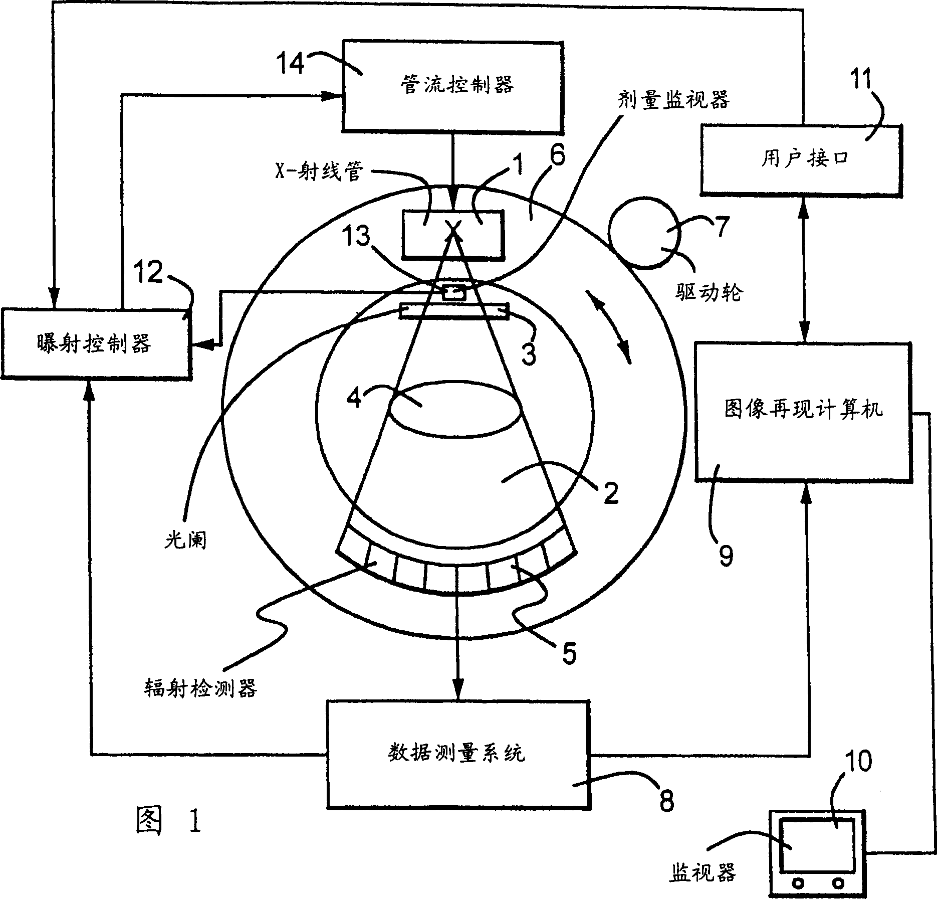
[0019] The computer scanning system shown in Fig. 1 has an X-ray tube 1 which emits an X-ray beam 2 from a focal point. The X-ray beam 2 is gated by a diaphragm 3 and passes through an examination subject 4 so as to be incident on a radiation detector 5 . Accordingly, the X-rays incident on the radiation detector 5 are attenuated by the examination subject 4, and the radiation detector 5 generates electrical signals corresponding to the attenuated X-rays incident thereon.

[0020] In the embodiment shown in FIG. 1 , both the X-ray tube 1 and the radiation detector 5 are mounted on a rotatable gantry 6 rotated by drive wheels 7 . The X-ray beam 2 is thus caused to rotate around the examination subject 4, making a series of perspectives obtained respectively at different perspective angles. Each perspective has associated with it a dataset of the aforementioned electrical signals. From the radiation detector 5, these data sets are provided for each fluoroscopy to a data measur...


PUM


Abstract

In a computed tomography apparatus and a method for automatic exposure control in computed tomography scanning, an exposure controller, during a first half of a revolution of an X-ray focus around a subject, calculates an actual attenuation profile of a slice of the subject from electrical signals generated by the radiation detector in the first half of the revolution, and calculates an extrapolated attenuation profile of the slice for a second half of the revolution from the actual attenuation profile, and adjusts an operating parameter of the X-ray source, such as the tube current, to modify the radiation dose emitted by the X-ray source during the second half of the revolution dependent on the extrapolated attenuation profile. The extrapolated attenuation profile can be calculated to achieve a predetermined target pixel noise in the image for the slice currently being scanned. In a spiral scan composed of a number of such slices, respectively different target pixel noise values can be used to appropriately set a radiation dose in the scan of each slice dependent on the radiation absorption of the slice of the subject currently being scanned.

Description

technical field [0001] The present invention relates to a method of automatically controlling exposure during a CT (computed tomography) scan, and a device for automatically controlling exposure in a CT scan. Background technique [0002] Previous generations of computed tomography systems have enhanced their ability to perform extensive scans over important areas of the subject's body. This capability improves the efficiency of CT equipment by reducing examination time and thereby increasing patient throughput. During a relatively long examination, eg a helical (spiral) examination covering a large part of the subject's body, the radiation absorbed by the patient varies significantly along the helical path. If no additional measures are taken, a CT system will use X-rays of constant intensity over the entire scan volume. For example, when a patient's shoulder-thoracic region is being scanned, the radiation absorbed by the patient in the shoulder slices is high, so high x-...

Claims


Application Information

Patent Timeline
IPC IPC(8): H05G1/44A61B6/03H05G1/34
CPCA61B6/544A61B6/542H05G1/34A61B6/027A61B6/032
Inventor 迪特马·亨切尔斯蒂芬·波普斯库克里斯托弗·苏斯海科·沃尔夫
Owner SIEMENS AG
Why Eureka
Try Eureka