Method and system for airway measurement

A technique of trachea, measured values, used in the field of methods and systems

Inactive Publication Date: 2004-06-09
GENERAL ELECTRIC CO
View PDF0 Cites 6 Cited by
  • Summary
  • Abstract
  • Description
  • Claims
  • Application Information

AI Technical Summary

Problems solved by technology

In other diseases, such as emphysema, it is difficult for fluoroscopy specialists to discern the extent of the disease just by looking at CT images

Method used


Image

  • Method and system for airway measurement
  • Method and system for airway measurement

Examples


Embodiment Construction

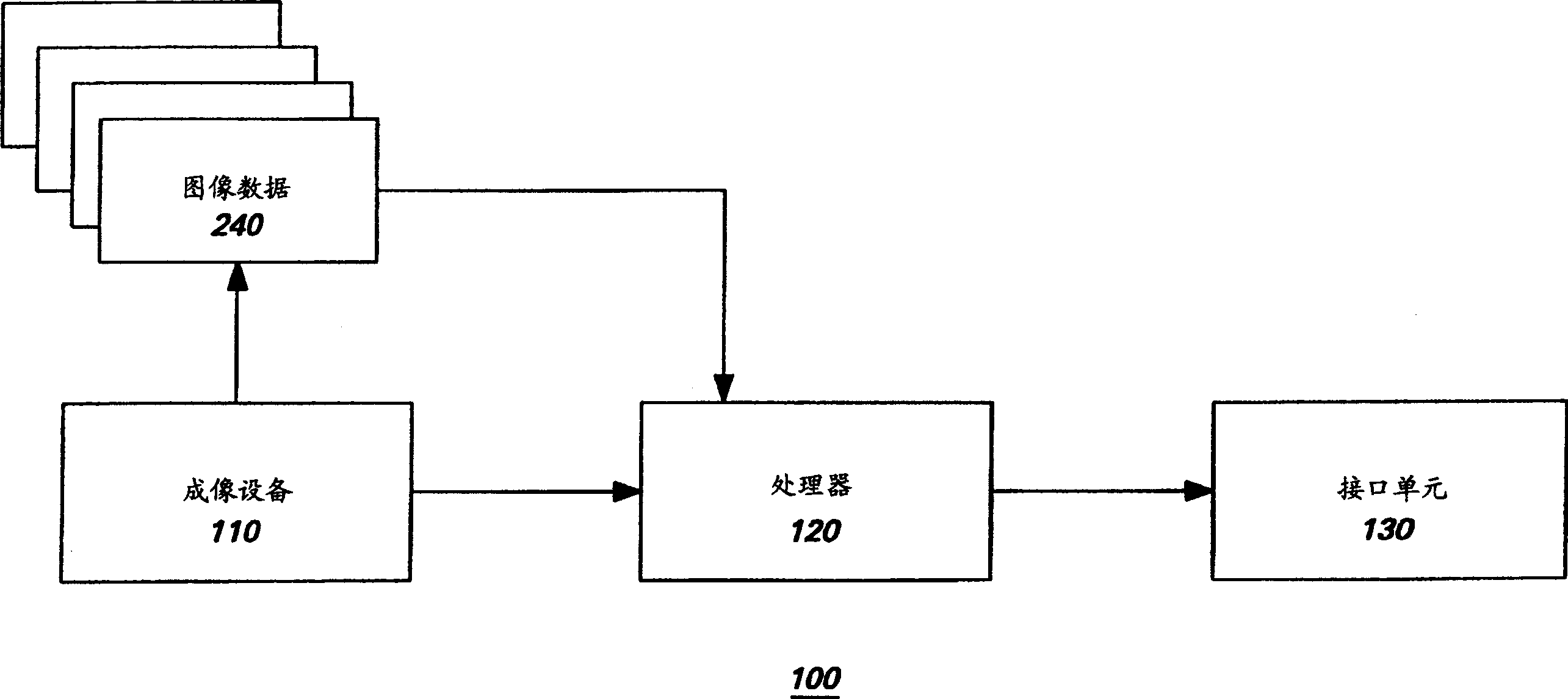
[0015] refer to figure 1 , shows a general block diagram of the system 100 for disease detection. The system 100 includes an imaging device 110, which may be selected from some prior art medical imaging devices, for generating several images. Typically, computed tomography (CT) and magnetic resonance imaging (MRI) systems are used to generate several medical images.

[0016] During CT imaging, a patient is placed in an imaging device and exposed to several X-rays measured by a series of X-ray detectors. A single X-ray beam traverses patient-specific thin sections, or "slices." The detector measures the amount of transmitted radiation. This information is used to calculate the X-ray attenuation coefficients for the sampling points in the body. A grayscale image is then constructed based on the calculated X-ray attenuation coefficients. Shades of gray in the image represent the amount of X-ray absorption at points within the slice. Slices obtained during a CT procedure can...


PUM


Abstract

A method for measuring tubular anatomical structures, for example lung airways, comprises isolating by at least one segmentation process a given tubular anatomical structure of interest and measuring at least one attribute of the structure of interest. A system for measuring lung airways using acquired image data is provided. The system comprises an imaging device for acquiring the image data and an image processing device coupled to the imaging device. The imaging processing device is configured for isolating by at least one segmentation process a given airway of interest, fitting an inner ellipse to an inner boundary of the given airway and an outer ellipse to an outer boundary of the airway structure using statistical techniques at a given point in the airway, and further configured for generating measurements of the given airway using the inner and outer ellipses.

Description

technical field [0001] The present invention relates to a method and system for processing medical image data to assist in the detection and diagnosis of disease, and more particularly to a method and system for detecting lung disease from medical images obtained from X-ray computed tomography (CT) systems. Background technique [0002] Radiograph systems are more commonly used diagnostic tools for the purpose of detecting lung disease in humans. Lung diseases such as bronchitis, emphysema, and lung cancer can also be detected on chest X-ray and CT. However, CT systems typically provide more than 80 discrete images for a single CT scan, thereby providing a fluoroscopy specialist with a considerable amount of information to interpret the images and detect suspicious areas that may indicate disease. [0003] Suspect areas are defined as areas for which a trained fluoroscopy specialist would recommend follow-up diagnostic imaging, biopsy, pulmonary function testing, or other m...

Claims


Application Information

Patent Timeline
Patent Type & Authority Applications(China)
IPC IPC(8): G01R33/32A61B5/055A61B6/03A61B8/00G06T1/00G06T7/00G06T7/60
CPCG06T7/602G06T7/62
Inventor 詹姆斯·V·米勒马修·W·图雷克约瑟夫·L·芒迪彼得·H·图
Owner GENERAL ELECTRIC CO
Why Eureka
Try Eureka