Supercharge Your Innovation With Domain-Expert AI Agents!

X-ray imaging apparatus and method for moving an X-ray detector

A technology of a diagnosis device and a control method, which is applied in radiation safety devices, instruments for radiation diagnosis, diagnosis, etc., can solve the problems of stopping the X-ray detection part, uneven electromagnetic field, and complicated correction methods.

Inactive Publication Date: 2005-04-13
TOSHIBA MEDICAL SYST CORP
View PDF1 Cites 5 Cited by
  • Summary
  • Abstract
  • Description
  • Claims
  • Application Information

AI Technical Summary

Problems solved by technology

However, the correction method for this method is complicated, and it is difficult to perform stable correction of the capacitance value.
Moreover, the capacitance sensor in this method is installed around the X-ray detection section, so the formed electromagnetic field is uneven relative to the patient's body surface, especially when the patient's shape is convex, it cannot Correctly detect the distance between the X-ray detection unit and the closest patient part
[0012] Moreover, this method cannot correct other error factors other than the shape of the patient, so it is difficult to stop the X-ray detector at the desired position.

Method used


Image

  • X-ray imaging apparatus and method for moving an X-ray detector
  • X-ray imaging apparatus and method for moving an X-ray detector
  • X-ray imaging apparatus and method for moving an X-ray detector

Examples


Embodiment Construction

[0027] Embodiments of the present invention will be described below with reference to the drawings.

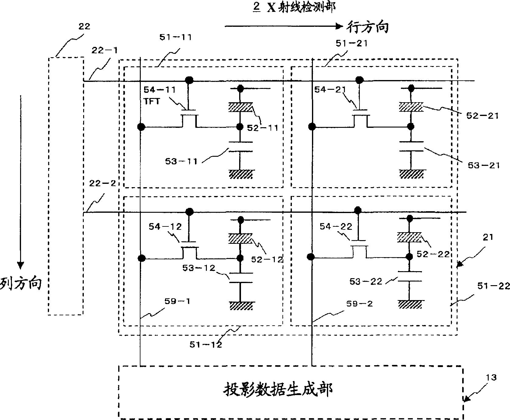
[0028] In the first embodiment, the front surface of the X-ray detection unit is covered with a thin plate-shaped capacitance sensor.

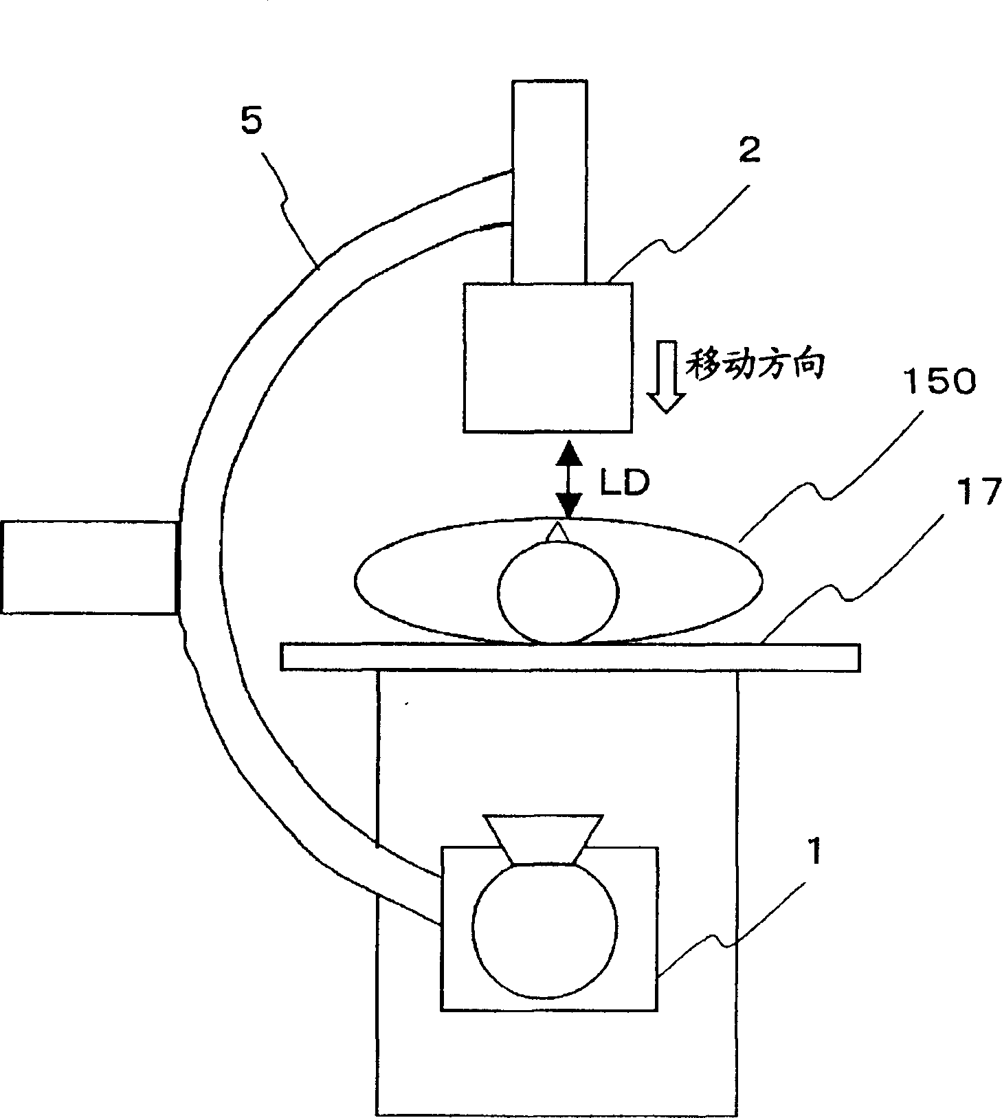
[0029]Also in this embodiment, when estimating the distance between the front of the X-ray detection unit and the patient's body surface, it is possible to use the electrostatic capacity correction set in advance with respect to the patient's shape, age, sex, obesity, etc. value, the value of the detected electrostatic capacitance is corrected, and the distance between the X-ray detection unit and the patient is estimated based on the corrected electrostatic capacitance.

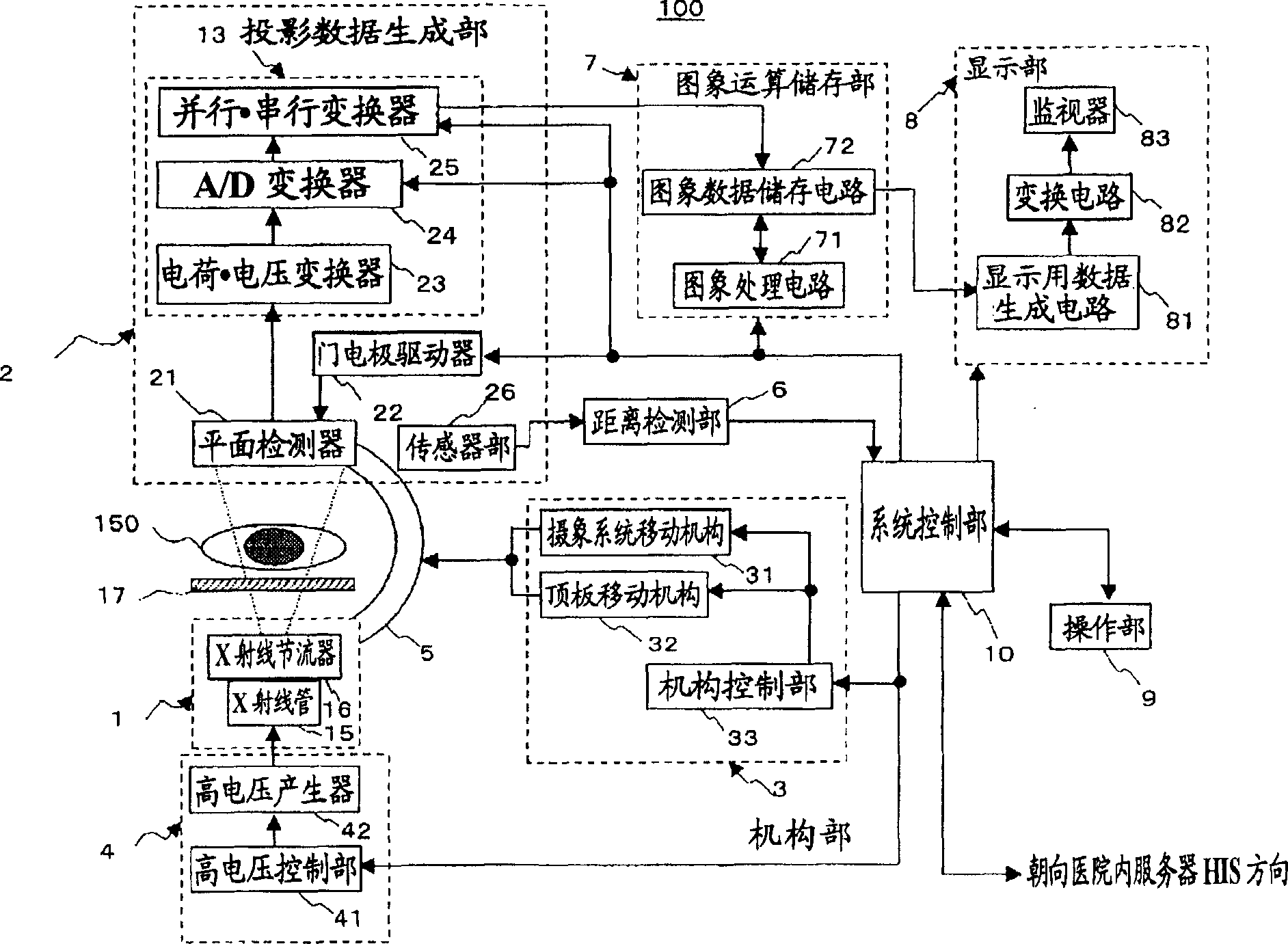
[0030] Refer below Figure 1 to Figure 5 , the configuration of the X-ray diagnostic apparatus as an embodiment of the present invention will be described. figure 1 It is a schematic block diagram showing the overall configuration of the X-ray diagnostic apparatus.

[003...


PUM


Abstract

An X-ray diagnostic device, characterized in that it can have an X-ray generating unit for irradiating X-rays to a subject; an X-ray detecting unit for detecting X-rays irradiated by the X-ray generating unit; an image data generation unit for generating X-ray image data at the imaging position from the X-ray information obtained by the X-ray detection unit; and moving the X-ray detection unit relative to the subject a mobile unit for use; an electrostatic capacity detection unit for detecting the electrostatic capacity at the X-ray detection unit using electrodes provided at least a part of the X-ray incident surface of the X-ray detection unit; and according to the A distance estimating unit for estimating the distance between the X-ray detecting unit and the subject based on the detected electrostatic capacity.

Description

technical field [0001] The invention relates to an X-ray diagnostic device and a movement control method of an imaging system. Background technique [0002] An X-ray diagnostic apparatus for circulatory organ diagnosis generally includes an X-ray generating unit and an X-ray detecting unit, a holding mechanism for holding these components, a diagnostic bed, and a signal processing unit. The holding mechanism adopts such as C-arm or Ω-arm, so that X-ray imaging can be performed at the best position and angle relative to the patient. [0003] As a detector used at the X-ray detecting portion of the X-ray diagnostic apparatus, X-ray film and I.I. (imaging·multiplier) are generally used. For the X-ray imaging method using this I.I., X-rays can be irradiated to the patient through the X-ray tube of the X-ray generating part, and the X-ray image information obtained through the patient can be converted into an I.I. optical image by I.I. elephant. This optical image can be trans...

Claims


Application Information

Patent Timeline
IPC IPC(8): G01B7/00A61B6/00A61B6/10
CPCA61B6/102G01B7/023
Inventor 太田聪
Owner TOSHIBA MEDICAL SYST CORP
Features
  • R&D
  • Intellectual Property
  • Life Sciences
  • Materials
  • Tech Scout
  • Unparalleled Data Quality
  • Higher Quality Content
  • 60% Fewer Hallucinations
Learn More