Animal model for toxicology and dose prediction

An animal, vertebrate technology, applied in the field of cell biology, can solve different problems

Inactive Publication Date: 2005-08-17
RAVEN BIOTECHNOLOGIES INC
View PDF3 Cites 4 Cited by
  • Summary
  • Abstract
  • Description
  • Claims
  • Application Information

AI Technical Summary

Problems solved by technology

This surgically removed tissue is more relevant to living tissue than autopsy tissue, but because of its proximity to diseased tissue and / or treatments the patient has undergone, the tissue may also be significantly different from normal tissue

Method used


Image

  • Animal model for toxicology and dose prediction
  • Animal model for toxicology and dose prediction
  • Animal model for toxicology and dose prediction

Examples


Embodiment 1

[0083] The generation of embodiment 1 non-human animal model

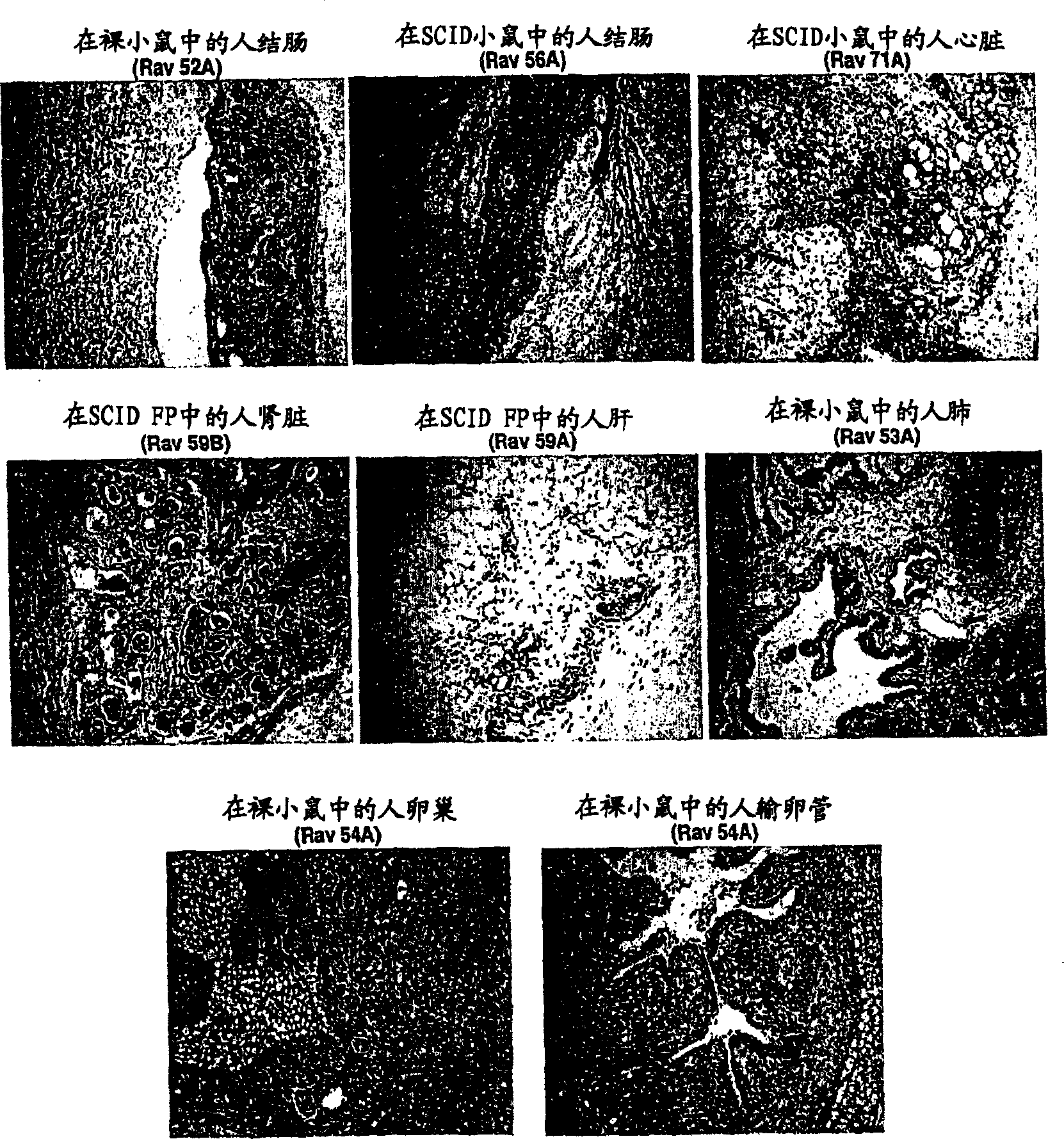
[0084] Tissues from normal embryonic organs (colon, heart, kidney, liver, lung, ovary, and fallopian tube) were cut into 1 mm cubed pieces and placed in the kidneys of nude mice (nu / nu) or SCID immunocompromised mice. in the bursa or fat pad. These tissues are left in the animals for 6-40 weeks to allow time to develop into mature tissues. Animals were euthanized and tissues were removed and sectioned for H&E staining and immunohistochemical evaluation.

[0085] figure 1 Results are shown for a series of implants in which tissues were allowed to mature for 4 months. In this example, for all references to "pictures" see figure 1 . Pictures 1, 2, 3, 6, 7 and 8 show implantation under the renal capsule and pictures 4 and 5 show implantation under the fat pad. Pictures 1, 6, 7 and 8 show the implantation of normal embryonic organs in nude (nu / nu) mice, while pictures 2, 3, 4 and 5 show the implantation of normal ...

Embodiment 2

[0086] Example 2 Mature Tissues for Safety / Efficacy Models

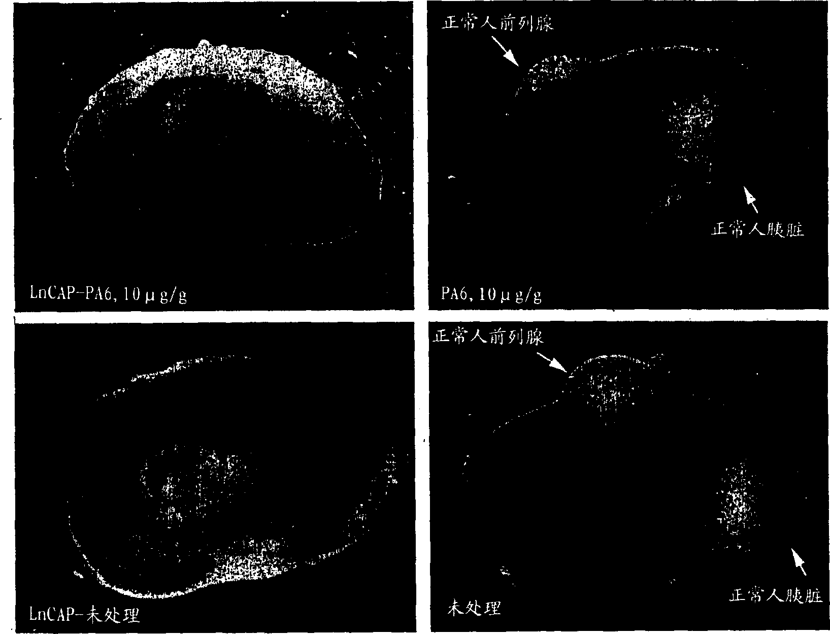
[0087] Small pieces of normal human prostate and pancreas were placed under the renal capsule and allowed to mature for 6 weeks. At this point, human prostate cancer cells (LnCAP) were placed under the contralateral renal capsule of the same animals and allowed to grow for an additional week. On day 7 after LnCAP tumor implantation, one animal was treated with 10 μg / g PA6 antibody (anti-human EpCAM) by intraperitoneal injection. Control animals were treated with saline injections. 4 injections in two weeks. At the end of this time, animals were euthanized and examined for tumor and normal tissue xenografts. animal kidneys figure 2 displayed in . figure 2 The left side shows LpCAP tumors and the right side shows normal tissue (a total of 9 weeks of development in this animal). The upper pictures are from treated animals and the lower pictures are from control animals. Other treated animals contained normal co...

Embodiment 3

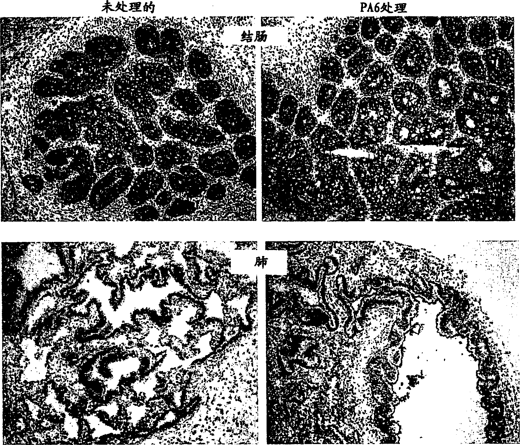
[0088] Example 3 Immunohistochemistry of Human Prostate and Human Colon Mature Tissues

[0089] Immunohistochemistry of human prostate and human colon mature tissues from experiments in figure 2 described in. While tumors were affected by antibody treatment leading to cell death and hemorrhage, normal tissues were not (A-D). To determine whether the tissue contained the antibody target (EpCAM), the tissue was stained with a directly labeled PA6 (anti-human EpCAM) antibody. Both treated and untreated tissues showed antibody binding. Mature human prostate tissue also stains strongly for prostate-specific antigen (PSA), a marker of prostate cells.


PUM

PropertyMeasurementUnit
molecular weightaaaaaaaaaa

Abstract

The invention relates to the use of fetal tissues to generate a tissue model in a non-human animal. The tissue model comprises target tissues allowed to progress through development in vivo in a non-human host in order to obtain tissues having a mature phenotype that can be sued to assess toxicity and / or efficacy of an agent.

Description

[0001] Cross-references to related applications: [0002] This application claims the benefit of US Provisional Application Serial No. 60 / 384,715, filed May 30, 2002, which is incorporated by reference in its entirety. field of invention [0003] The present invention belongs to the field of cell biology. More specifically, it relates to the use of embryonic tissue from one species that has been allowed to develop in vivo in a host of another species to obtain a tissue with a mature phenotype that can be used to evaluate the activity and toxicity of an agent. Background of the invention [0004] The use of animal models is critical for the proper evaluation of the efficacy and safety of new drugs. When an Investigational New Drug Application (IND) is submitted, trials are required in two species, usually rodents (rabbits, rats, mice, hamsters, guinea pigs, etc.) and often primates. Rodents are less expensive but have the problem of being too evolutionarily distant from hum...

Claims


Application Information

Patent Timeline
Patent Type & Authority Applications(China)
IPC IPC(8): A01K67/027A01KA01K33/00A61K35/00A61K35/12A61K49/00C12N5/07C12N5/073C12N5/074G01N33/15
CPCA01K67/0271A61K49/0008A01K2227/105A01K2227/10A01K2227/30A01K2267/03A01K67/027A61K35/00
Inventor J·P·马瑟P·F·扬
Owner RAVEN BIOTECHNOLOGIES INC
Why Eureka
Try Eureka