Method of fast increasing glandular related virus mediating gene in expression of inside of retina cell
A retinal cell and adenovirus technology, used in gene therapy, genetic engineering, plant genetic improvement, etc., can solve problems such as disease-causing retinal cells and degeneration
- Summary
- Abstract
- Description
- Claims
- Application Information
AI Technical Summary
Problems solved by technology
Method used
Image
Examples
Embodiment 1
[0069] Wild-type adenovirus accelerates and increases the transduction efficiency and gene expression level of AAV to retinal cells, but at the same time causes retinal cell lesions
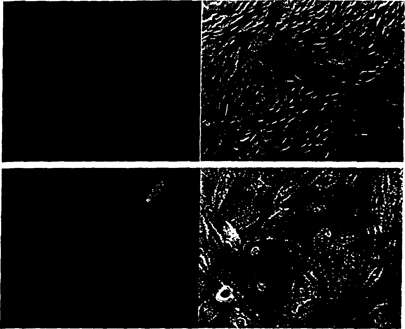
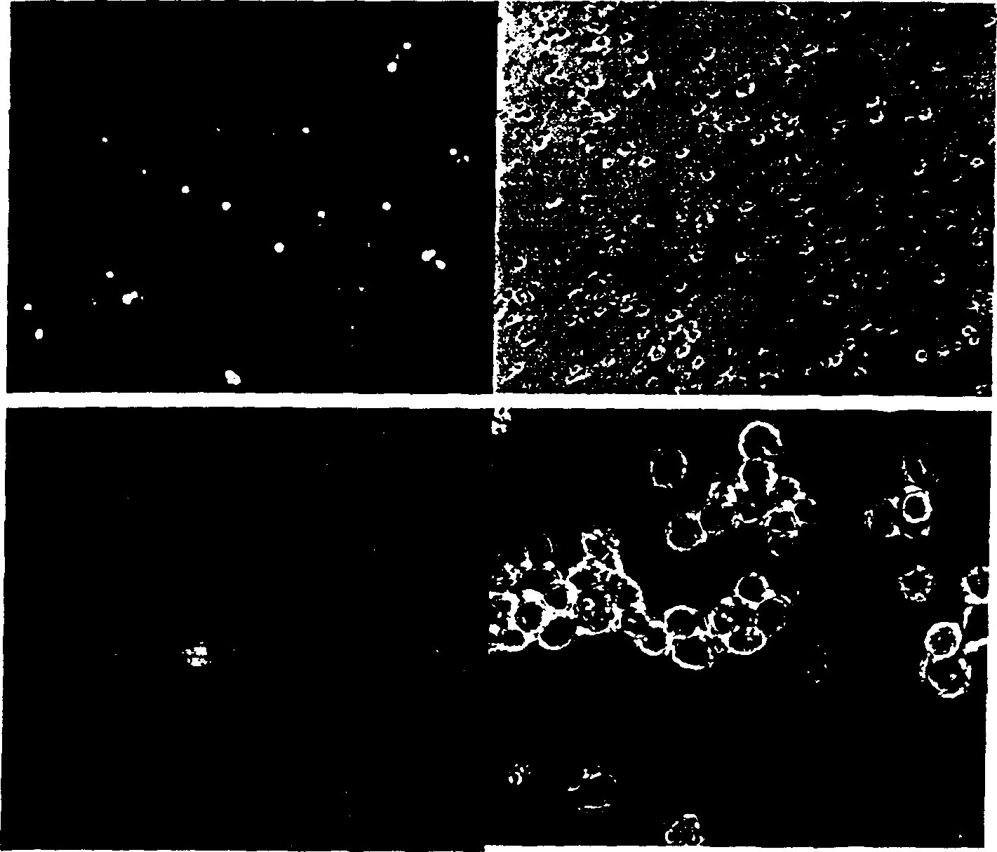
[0070] 2×10 per hole 5 RPE (APRE-19, ATCC) cells were seeded in 6-well cell culture plates, and after 24 hours, the cells adhered to the wall, and 1×10 8 or 1×10 9 AAV 2 -GFP with 1 x 10 3 , 1×10 4 or 1×10 5 DL309 was added to the cultured cells, and then observed with a fluorescent microscope every day and photographed to record the brightness of GFP fluorescence, cell morphology and disease state of the infected cells. After 5-7 days, trypsinization was performed, the number of GFP-positive cells and GFP fluorescence intensity were detected by flow cytometry, and the copy number of GFP gene and the expression level of GFP were detected by PCR and Western blot.
[0071] The above test results show that when AAV 2 - The amount of GFP virus used was 1 × 10 8 When DL309 was not added, GFP e...
Embodiment 2
[0074] Conditionally replicating adenovirus accelerates and improves AAV transduction efficiency and gene expression level in retinal cells
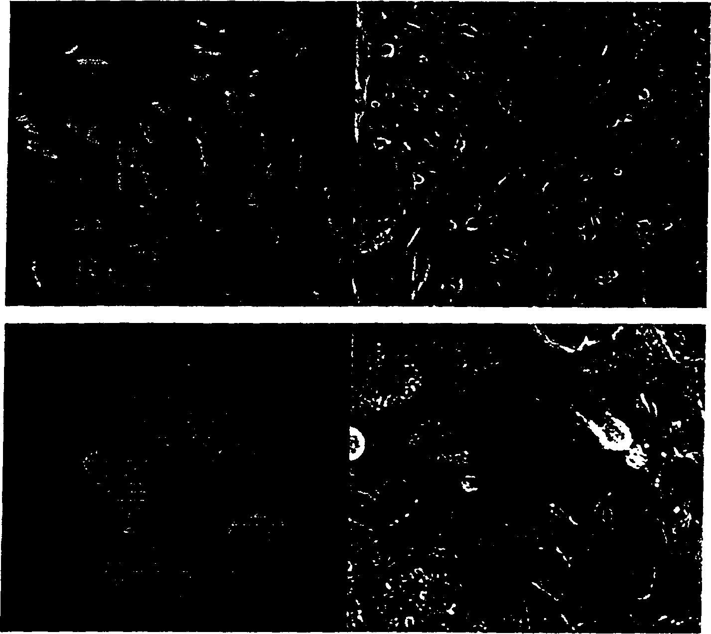
[0075] The conditional replication adenovirus is different from the wild-type adenovirus DL309 in that although it has E1A and E1B genes, its expression is regulated by the TERT gene promoter or the HSP gene promoter, respectively. Therefore, its replication is regulated to a certain extent, with better safety and less toxic and side effects. The dosage of Ad-TERT-E1A / E1B and Ad-HSP-E1A / E1B in the in vitro experiment was 1×10 3 -1×10 6 pfu. AAV 2 -The amount of GFP was 1×10 8 and 1×10 9 Viral particles; for each RPE cell, the amount of adeno-associated virus is about 5 × 10 2 -5×10 3 virus particles, the amount of conditionally replicating adenovirus is about 0.005-5 pfu. The ratio of conditionally replicating adenovirus to adeno-associated virus was 1:1×10 2 -1:1×10 5 ; the dosage of each nerve cell is roughly 10 4 -10 5 For...
Embodiment 3
[0083] Replication-defective adenovirus accelerates and increases AAV transduction efficiency and gene expression levels in retinal cells
[0084] The amount of Ad-Luc used in the in vitro experiment was 1×10 3 , 1×10 4 , 1×10 5 and 1×10 6 pfu, AAV 2 The dosage is 1×10 8 or 1×10 9 virus particles. The same design as the conditional replicating adenovirus experiment, for each RPE cell, the amount of adeno-associated virus is about 5×10 2 -5×10 3 For each virus particle, the amount of replication-defective adenovirus Ad-Luc is about 0.005-5pfu; the ratio of Ad-Luc to adeno-associated virus is 1:10 2 -1:10 6 ; the dosage of each nerve cell is roughly 10 4 -10 5 For each virus particle, the dosage of Ad-Luc is about 0.05-5pfu. The ratio of Ad-Luc to Adeno-associated virus was 1:10 2 -1:10 5 .
[0085] 2×10 per hole 5 RPE (APRE-19, ATCC) cells were seeded in 6-well cell culture plates, and after 24 hours, the cells adhered to the wall, and 1×10 8 or 1×10 9 AAV 2 ...
PUM
 Abstract
Description
Claims
Application Information
 - R&D
- Intellectual Property
- Life Sciences
- Materials
- Tech Scout
- Unparalleled Data Quality
- Higher Quality Content
- 60% Fewer Hallucinations
Browse by: Latest US Patents, China's latest patents, Technical Efficacy Thesaurus, Application Domain, Technology Topic, Popular Technical Reports.
© 2025 PatSnap. All rights reserved.Legal|Privacy policy|Modern Slavery Act Transparency Statement|Sitemap|About US| Contact US: help@patsnap.com


