Method and system for computed tomography illustration of the movement of a heart

A tomography, computer technology, applied in computer tomography scanners, instruments for radiological diagnosis, diagnosis, etc., can solve the problems of cell DNA damage, increase the risk of oncomycin, injury, etc., and achieve the effect of reducing radiation dose.

Inactive Publication Date: 2006-11-01
SIEMENS AG
View PDF0 Cites 4 Cited by
  • Summary
  • Abstract
  • Description
  • Claims
  • Application Information

AI Technical Summary

Problems solved by technology

[0004] The problem with such heart scans is that, on the one hand, any avoidable radiation dose to the patient is avoided, since even small residual radiation doses can produce damage to the cellular DNA, which sometimes leads to elevated cancer risk. On the other hand, the contrast agent used also has a harmful effect, so here also efforts should be made to make the administration of the contrast agent as small as possible.

Method used


Image

  • Method and system for computed tomography illustration of the movement of a heart
  • Method and system for computed tomography illustration of the movement of a heart
  • Method and system for computed tomography illustration of the movement of a heart

Examples


Embodiment Construction

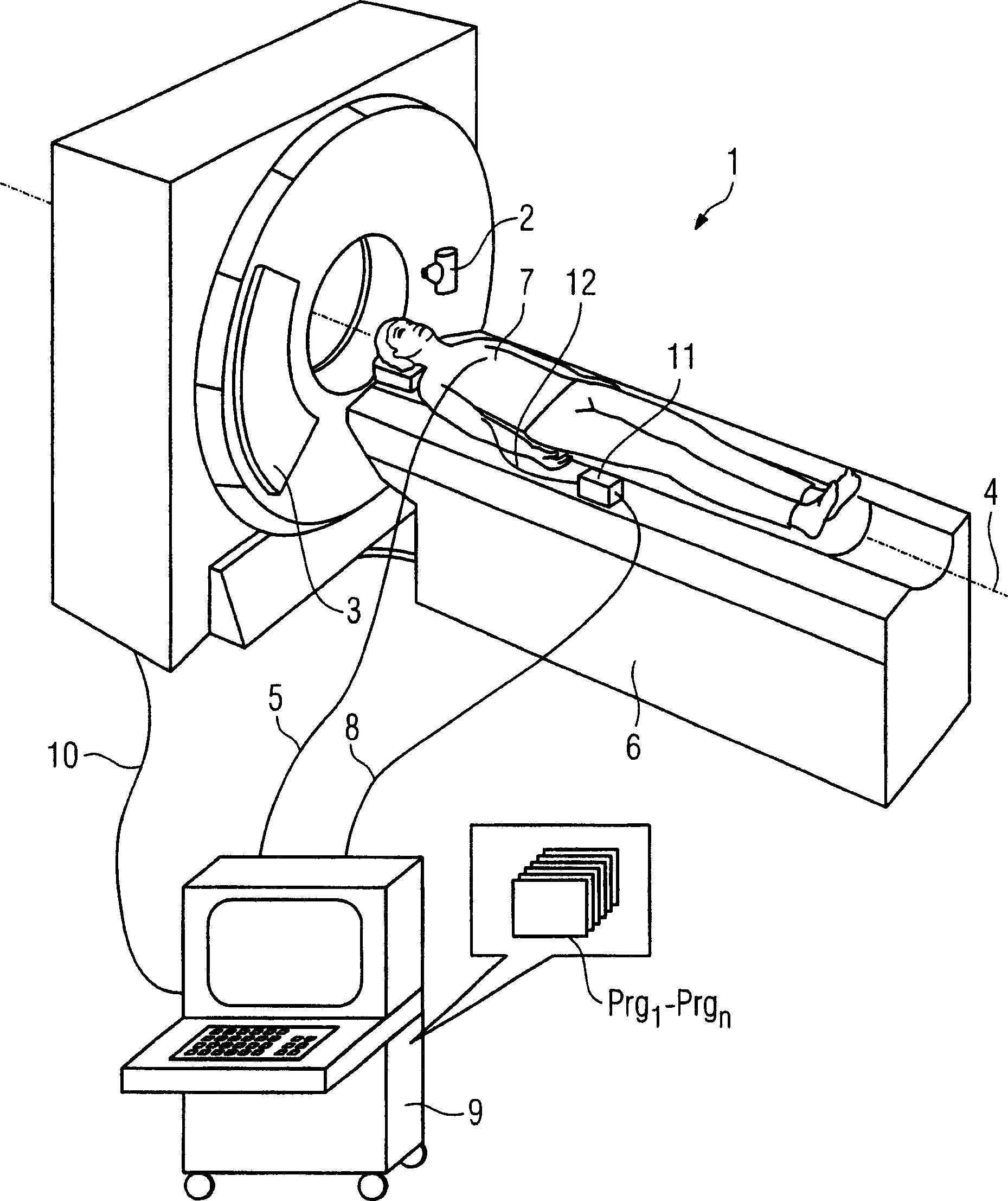
[0036] figure 1 Shown is a system 1 according to the invention comprising a control and computing unit 9, in which a program Prg is stored in a data memory 1 -Prg nAnd the control and data processing of the whole system are carried out according to the present invention. It is of course also possible within the scope of the invention to distribute individual method steps and control tasks on different computers. The control and computing unit 9 shown here is connected via control and data lines 10 to the actual CT system, which has a gantry-mounted x-ray tube 2 moving on a circular path for scanning a patient and A detector set opposite it. During the scanning process, a patient 7 on a patient couch 6 movable in the direction of the system axis 4 is moved forward in the direction of the system axis 4 , so that a helical scan relative to the patient coordinate system is finally performed. The system 1 according to the invention also has an EKG integrated in the computing un...


PUM


Abstract

A method and a system are disclosed for computed tomography illustration of the movement of a heart in the cardiac cycle with the aid of a spiral CT. In the method, a patient is administered a contrast medium via an electronically controllable apparatus. A stationary prescan of a cardiac artery is carried out in order to determine the sufficient filling of the artery with the automatically applied contrast medium. When a sufficient contrast medium filling is detected, the current heart rate of the heart being examined is measured, and a maximum possible feed rate for a spiral scan and a duration of the spiral scan are determined on the basis of the current heart rate. Subsequently, the spiral scan is carried out over the heart region with the maximum possible feed rate.

Description

technical field [0001] The invention relates to a method and a system for computer tomographic visualization of cardiac motion using helical CT, in which a 3D image series of the heart cycle is generated by means of a helical scan. Background technique [0002] Similar methods are generally known. Typically, a helical scan of the heart region is performed in order to generate an image series of the heart movement during the cardiac cycle, wherein the heart is completely scanned in all cycle phases and the time course of the cycle is reproduced in a stereoscopic display from at least one cardiac cycle. An EKG (electrocardiogram) is usually used for synchronizing the recorded image data, but it is also possible to determine the motion of the heart solely from the detector output data of the computed tomography and thus assign the recorded detector data to the individual cardiac cycle phases , thus integrating into a complete record of the heart over a typical cardiac cycle. ...

Claims


Application Information

Patent Timeline
Patent Type & Authority Applications(China)
IPC IPC(8): A61B6/03
CPCA61B6/032A61B6/027A61B6/481A61B6/488A61B6/503A61B6/541
Inventor 迪特尔·波音阿克塞尔·库特纳约翰·尤伯勒
Owner SIEMENS AG
Why Eureka
Try Eureka