X-ray digital imaging correction method

A correction method and digital imaging technology, applied in the field of X-ray digital imaging correction, can solve the problems of incomplete X-ray response coefficients, uneven spatial distribution, affecting image quality, etc.

Inactive Publication Date: 2006-12-20
SHANGHAI MEDICAL EQUIPMENT WORKS CO LTD
View PDF0 Cites 28 Cited by
  • Summary
  • Abstract
  • Description
  • Claims
  • Application Information

AI Technical Summary

Problems solved by technology

[0006] 1) Although the X-ray response of a single pixel of the detector is linear within the linear exposure dose range, the X-ray response coefficients of different pixels are not completely consistent, resulting in uneven images
[0007] 2) Image unevenness caused by inconsistencies in peripheral circuits such as row drive circuits, read amplifiers, and A / D converters
[0008] 3) Image inhomogeneity caused by the inherent spatial inhomogeneity of the incident X-ray itself
[0011] Based on a systematic analysis of the imaging chain, it is known that there are inherent defects in the detector panel, which, if not systematically corrected, will affect the final image quality

Method used


Image

  • X-ray digital imaging correction method
  • X-ray digital imaging correction method
  • X-ray digital imaging correction method

Examples


Embodiment Construction

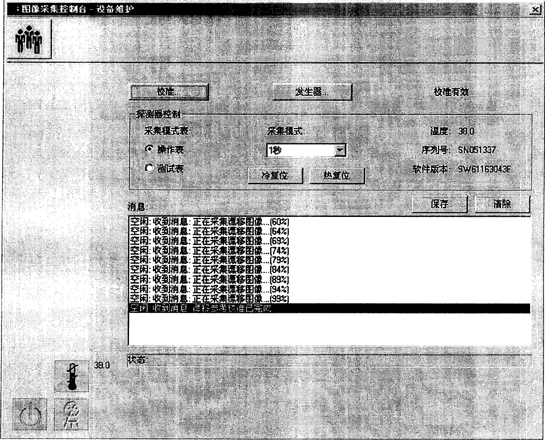
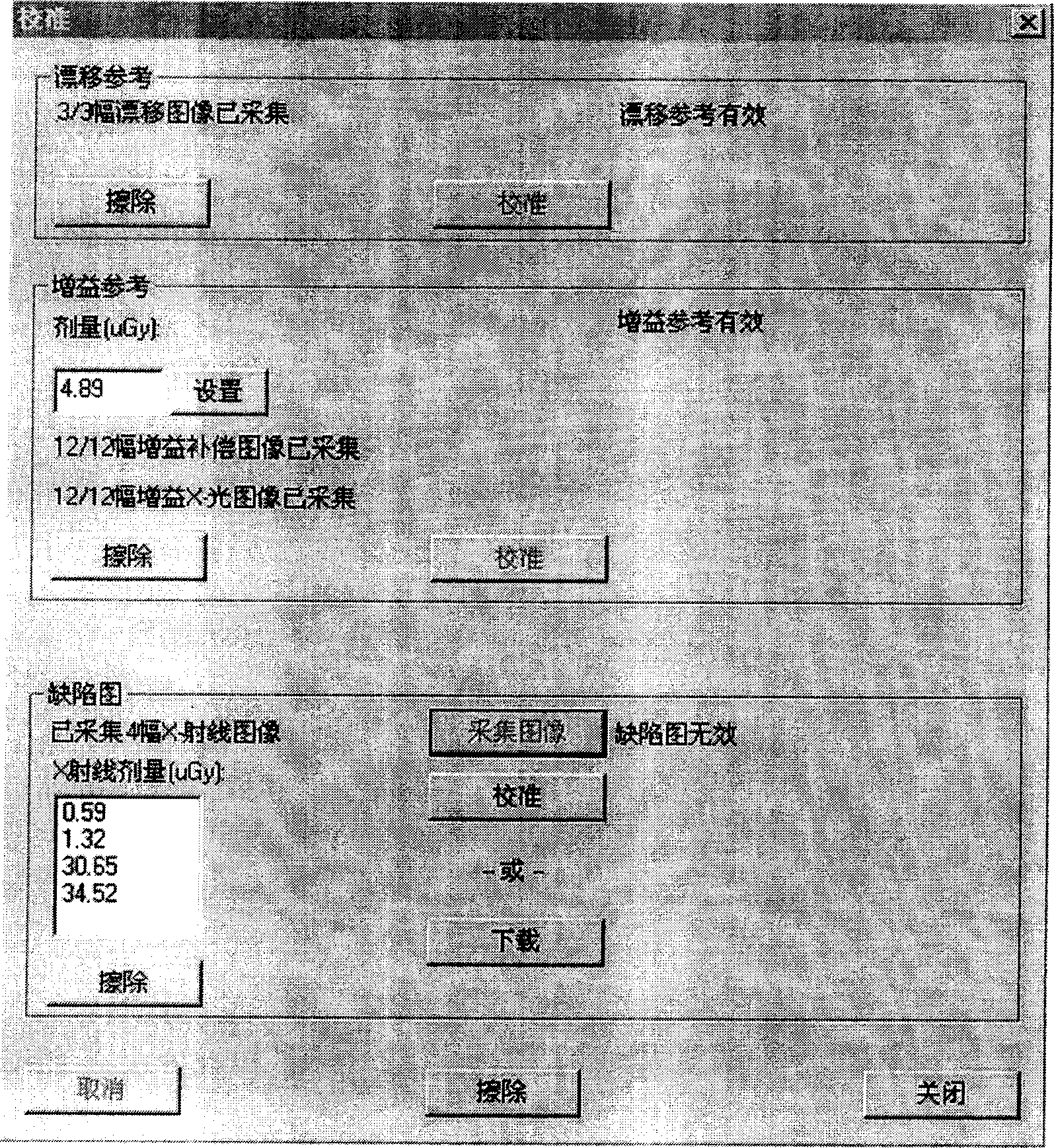
[0047] As shown in accompanying drawing 6-7, the present invention provides a kind of correction method of X-ray digital imaging, and it comprises the following steps:

[0048] 1. Start to collect image data and get the original image;

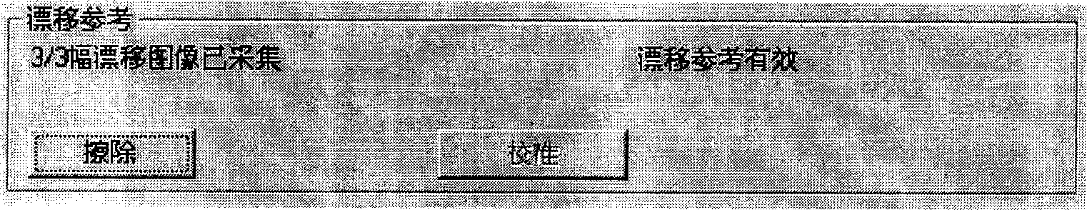
[0049] 2. Perform drift correction on the above original image;

[0050] 3. Perform spatial non-uniformity correction on the above original image;

[0051] 4. Perform dead point correction on the above original image;

[0052] 5. The final clean image can be used for image storage and expression.

[0053] The flat panel detector used in the embodiment of the present invention adopts the Pixium 4600 type flat panel detector provided by Trixell Company to calibrate. The external environment is in a constant temperature operation room. After the detector is powered on for 4 hours, the temperature of the detector is basically stable. . The distance from the radiation source to the SID of the flat panel is 150cm, a 21mm aluminum filter is used,...


PUM


Abstract

The invention discloses a correcting method of X-ray digital imaging in the digital imaging technological domain, which comprises the following steps: utilizing mathematical model to correct shift of digital flat according to physics analysis of imaging chain; correcting spatial unevenness and broken points to obtain stable, entire and correct digital image of X-ray information; fitting for reserving and expressing image. The invention can correct systemic defect of direct digital intrinsic imaging system through predisposal system, which improves imaging effect.

Description

technical field [0001] The invention relates to the technical field of digital imaging, in particular to a correction method for X-ray digital imaging. Background technique [0002] Flat-panel detectors have been introduced into the field of medical X-ray in recent years. X-ray is converted into visible light by CsI:Tl scintillator, and then converted into charge by a-Si photodiode. The charges are then amplified and digitized for readout, allowing high-quality images to be obtained. Most of the detectors currently used in digital imaging systems in the field of medical X-ray are this type of detectors. [0003] Since the direct digital imaging system uses a large-scale solid-state detector array as the image acquisition component, it is inevitable to encounter solid-state detection such as drift (offset), spatial non-uniformity (Gain), bad point (defect point), and nonlinear response. How to properly correct the inherent defects of the sensor array has become a very impor...

Claims


Application Information

Patent Timeline
Patent Type & Authority Applications(China)
IPC IPC(8): G03B42/02
Inventor 杨叶
Owner SHANGHAI MEDICAL EQUIPMENT WORKS CO LTD
Why Eureka
Try Eureka