System and method for three-dimensional reconstruction of a tubular organ

A technology for three-dimensional reconstruction of tubular organs, applied in the field of medical imaging systems for angiography, and can solve problems such as incorrect results, adequate support for coronary angiography, and trouble.

Inactive Publication Date: 2006-12-20
PAIEON INC
View PDF12 Cites 22 Cited by
  • Summary
  • Abstract
  • Description
  • Claims
  • Application Information

AI Technical Summary

Problems solved by technology

[0018] Due to the aforementioned shortcomings of the prior art, current 2D QCA systems do not give adequate support for coronary angiography (for ex

Method used


Image

  • System and method for three-dimensional reconstruction of a tubular organ
  • System and method for three-dimensional reconstruction of a tubular organ
  • System and method for three-dimensional reconstruction of a tubular organ

Examples


no. 1 example

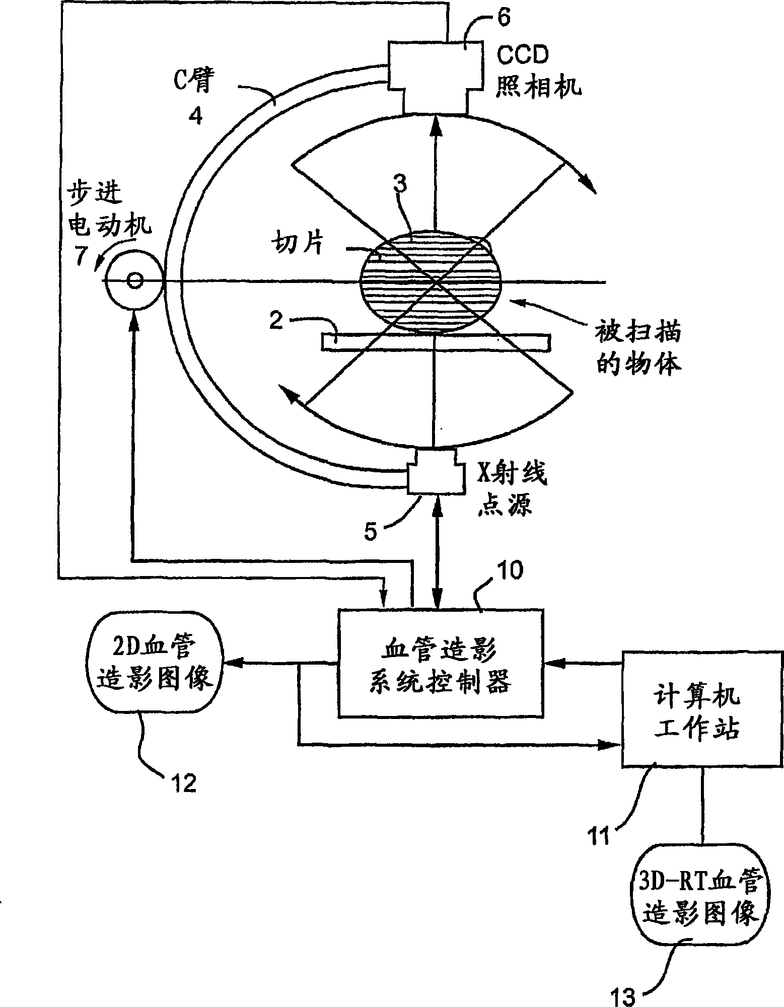
[0083] graphic gathering

[0084] During catheterization of a patient, a plurality of angiographic cine-projected two-dimensional (2D) x-ray images are captured substantially in real time and displayed on a monitor. In addition to these images, C-arm angulation data and ECG data can also be acquired. With the ECG sensor, an ECG gating process can be used to render the optimal ("best") image (end-diastolic frame) from each captured angiographic cine-projected image.

[0085] Capture of the movie show can be done in an analog fashion (eg with a frame grabber) or via a standard DICOM connection (preferred). DICOM is an acronym for "Digital Imaging and Communications in Medicine", and is a file format and digital communication protocol that allows medical equipment and software from different manufacturers to communicate with each other for easy sharing of medical data.

[0086] After image capture, the operator can perform catheter calibration on the image according to known me...

no. 2 example

[0174] It is an object of this group of embodiments of the invention to provide a method and system for three-dimensional reconstruction of tubular organs from angiographic projections. Specifically, the second group of embodiments improves the 3DR method of epipolar geometry conditions by providing additional considerations to the 3D reconstruction process, thereby providing precise correspondence between different projections, so that even in the presence of said geometric distortion and exopolar Accurate 3D models can also be provided under extremely problematic conditions.

[0175]According to a second set of embodiments, the proposed reconstruction method is based on the epipolar geometry enhanced by incorporating other considerations into the reconstruction process. These other considerations include, for example, parameters of the tubular organ derived from images along its centerline and local centerline directions, such as radius and densitometric (grayscale) values. ...

no. 3 example

[0244] It is an object of the third group of embodiments of the present invention to provide a method and system for three-dimensional organ reconstruction from more than two angiographic projections in an automated manner, i.e. without additional user interaction, i.e. Tubular organs are identified on radiographs.

[0245] Three-dimensional reconstruction of a tubular organ such as an artery from two projections can be achieved via methods known in the art. Typically, this requires user interaction to identify the organ of interest in the first two views. Once this reconstruction is available, a third group of embodiments provides a method for automatically updating based on two or more additional projections. When a 3D model reconstructed from two projections is available, it is projected onto an additional image plane according to the specific viewing geometry of this additional existing projection. This causes a large geometric distortion representing an unknown shift be...


PUM


Abstract

Embodiments of the present invention include methods and systems for three-dimensional reconstruction of a tubular organ (for example, coronary artery) using a plurality of two-dimensional images. Some of the embodiments may include displaying a first image of a vascular network, receiving input for identifying on the first image a vessel of interest, tracing the edges of the vessel of interest including eliminating false edges of objects visually adjacent to the vessel of interest, determining substantially precise radius and densitometry values along the vessel, displaying at least a second image of the vascular network, receiving input for identifying on the second image the vessel of interest, tracing the edges of the vessel of interest in the second image, including eliminating false edges of objects visually adjacent to the vessel of interest, determining substantially precise radius and densitometry values along the vessel in the second image, determining a three dimensional reconstruction of the vessel of interest and determining fused area (cross-section) measurements along the vessel and computing and presenting quantitative measurements, including, but not limited to, true length, percent narrowing (diameter and area), and the like.

Description

[0001] Priority claims and related applications [0002] This application requires U.S. Provisional Patent Application 60 / 505,430 filed September 25, 2003, U.S. Provisional Patent Application 60 / 506,178 filed September 29, 2003, and June 7, 2004 under 35 U.S.C. § 119(e) Priority to filed US Provisional Patent Application 60 / 577,981, the disclosure of which is hereby incorporated by reference in its entirety. technical field [0003] The present invention relates to medical imaging systems, in particular to medical imaging systems such as those used in angiography. Background technique [0004] Blood vessel stenosis (stenosis), such as arterial stenosis, refers to the narrowing of the inner lumen of an artery due to the formation of plaques on the inner wall of the artery. The severity of the narrowing depends on the amount of plaque obstruction in the cross-sectional area of ​​the lumen. Although arterial narrowing can occur in any artery of the body (for example, the caro...

Claims


Application Information

Patent Timeline
IPC IPC(8): G06K9/00
Inventor 摩舍·克雷曼迈克尔·扎克
Owner PAIEON INC
Why Eureka
Try Eureka