
Method for diagnosing inflammatory bowel disease

a technology for inflammatory bowel disease and bowel disease, which is applied in the direction of material testing goods, measurement devices, instruments, etc., can solve the problems of colonoscopy in ibd patients, long remission, and high cos

Active Publication Date: 2018-06-19
DIAGNODUS
View PDF5 Cites 0 Cited by
  • Summary
  • Abstract
  • Description
  • Claims
  • Application Information

AI Technical Summary

Benefits of technology

This approach provides a reliable, non-invasive means for diagnosing IBD, monitoring treatment effectiveness, and predicting disease relapse, with improved sensitivity and specificity compared to existing methods, facilitating more effective management of IBD.

Problems solved by technology

In most cases it is characterized by long remissions and incidental disabling flare-ups usually requiring treatment.
Differentiation between IBD and non-inflammatory diarrhoeas, such as Irritable Bowel Syndrome (IBS) constitutes another major problem, especially given that IBS affects 10-15% of the adult population in most developed Western countries (Agarwal & Whorwell, 2006; Khan & Chang, 2010).
IBD diagnosis confirmation usually requires colonoscopy, an efficient diagnostic procedure that is, however, highly invasive, expensive and can sometimes cause dangerous complications.
Repeated colonoscopies in IBD patients may be dangerous; therefore currently IBD activity and therapy efficiency are usually assessed by disease activity indexes based on severity of clinical manifestations and results of indirect laboratory analyses (Satsangi et al, 2006).
However, genetic approaches have not provided a clinically applicable method so far, being mostly confined to the prediction of Crohn's disease risk (Weersma et al, 2009) and assessment of pharmacogenetics of drugs used for its treatment (Roberts & Barclay, 2012).
In addition to limited diagnostic efficiency, most of these methods are exceedingly technically complex.
Although the outlined peripheral blood or serological marker panels may potentially be useful for differentiating UC from CD, disease monitoring and defining therapeutic strategies (Peyrin-Biroulet et al, 2007), they do not perform better than non-specific C-reactive protein (Palmon et al, 2008).
None of them is currently applied for practical clinical use.
Nevertheless, an attempt to use this marker for paediatric UC monitoring was not successful (Turner et al, 2010).
However, M2-PK is not applied in clinical practice.
Some authors also suggested that inflammation-related neutrophil degranulation can be detected in stool samples by quantifying myeloperoxidase (Wagner et al, 2008; Masoodi et al, 2011) and human neutrophil lipocalin (Nielsen et al, 1996, 1999), but these tests are not sufficiently studied to be proposed for IBD detection.
On the basis of the existing published evidence faecal EDN was not regarded as a promising biomarker of IBD, being less reliable than calprotectin or other stool biomarkers (Wagner et al, 2008; Foell et al, 2009).
This constitutes a serious obstacle for using it for diagnostic purposes.
The presented background information shows that despite the availability of a number of potentially promising biomarkers of intestinal inflammation the field is still poorly developed.
The only clinically employed biomarker-based test for IBD is stool calprotectin detection, the applicability of which is considerably limited due to the necessity of stool collection and handling.
Presently there is no reliable alternative non-invasive test for IBD.

Method used


Image

  • Method for diagnosing inflammatory bowel disease
  • Method for diagnosing inflammatory bowel disease
  • Method for diagnosing inflammatory bowel disease

Examples


Embodiment Construction

[0180]The present invention will now be further described. In the following passages, different aspects of the invention are defined in more detail. Each aspect so defined may be combined with any other aspect or aspects unless clearly indicated to the contrary. In particular, any feature indicated as being preferred or advantageous may be combined with any other feature or features being indicated as being preferred or advantageous.

[0181]As previously discussed there exists a need to develop new methods for the diagnosis of inflammatory bowel diseases. In particular, there exists a particular need to develop a non-invasive method for the detection of these diseases.

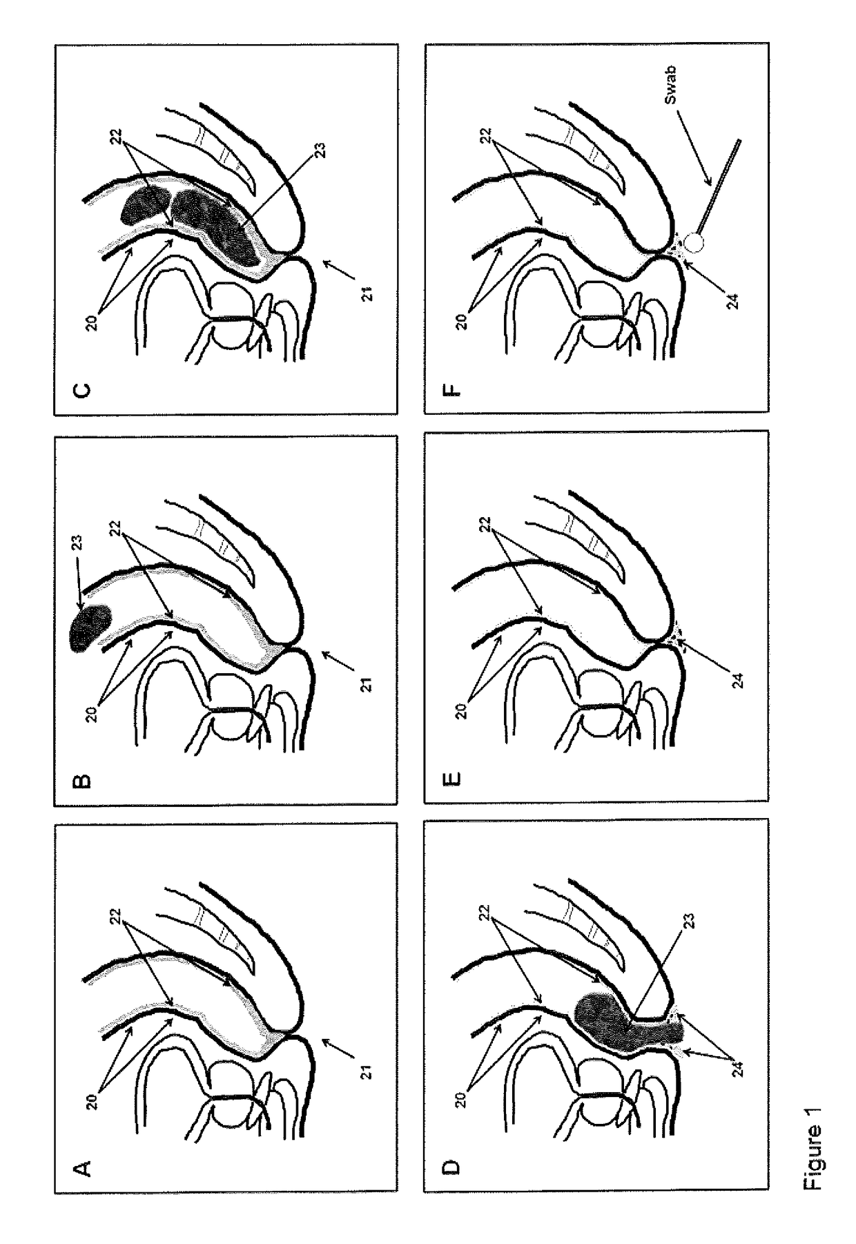
[0182]The inventors have surprisingly identified that a considerable portion of the colonic mucocellular layer is evacuated together with stool, and more importantly fragments of the mucocellular layer remain on the surface of the external anal / perianal area following defaectaion. Usually these fragments would be removed...


PUM

PropertyMeasurementUnit
concentrationaaaaaaaaaa

Abstract

The present invention relates to a method for diagnosing inflammatory bowel disease (IBD), the method comprising determining the concentration of at least one IBD-specific biomarker in a sample of the colonic mucocellular layer obtained from a subject.

Description

RELATED APPLICATIONS[0001]The present application is a U.S. National Stage application under 35 USC 371, claiming priority to International patent application Serial No. PCT / GB2013 / 052935, filed on Nov. 7, 2013. The present application is also a Continuation-in-Part of U.S. patent application Ser. No. 14 / 115,529, filed Jan. 21, 2014, which is a U.S. National Stage application under 35 USC 371, claiming priority to International patent application Serial No. PCT / GB2012 / 050964, filed on May 3, 2012, which claims priority from GB 1107466.3 filed on May 5, 2011. All of the aforementioned applications are incorporated herein by reference in their entirety.SEQUENCE LISTING[0002]The instant application contains a Sequence Listing which has been submitted in ASCII format via EFS-Web and is hereby incorporated by reference in its entirety. Said ASCII copy, created on Nov. 11, 2013, is named sequence listing 7.11.13 (4526563-1).txt and is 9 KB in size.FIELD OF THE INVENTION[0003]The present i...

Claims


Application Information

Patent Timeline
Patent Type & Authority Patents(United States)
IPC IPC(8): G01N33/68
CPCG01N33/6893G01N2800/065
Inventor LOKTIONOV, ALEXANDREBANDALETOVA, TATIANA
Owner DIAGNODUS
Learn More