Detection of stent struts relative to side branches

a technology of stent struts and side branches, applied in the field of system and method for stent detection, can solve the problems of difficult or impossible to detect strut shadows overlying side branches, struts over jailed side branches are difficult to detect, and stent flow-limiting stenoses are often presen

Active Publication Date: 2018-11-27
LIGHTLAB IMAGING
View PDF142 Cites 5 Cited by
  • Summary
  • Abstract
  • Description
  • Claims
  • Application Information

AI Technical Summary

Problems solved by technology

However, flow-limiting stenoses are often present in the vicinity of vascular side branches.
Since side branches are vital for carrying blood to downstream tissues, jailing can have an undesired ischemic impact and also can lead to thrombosis.
However, struts over jailed side branches are difficult to detect via this method.
As a result, it is difficult or impossible to detect strut shadows overlying side branches.
Consequently, jailing struts are easily missed by the shadow based detection methods.

Method used


Image

  • Detection of stent struts relative to side branches
  • Detection of stent struts relative to side branches
  • Detection of stent struts relative to side branches

Examples


Embodiment Construction

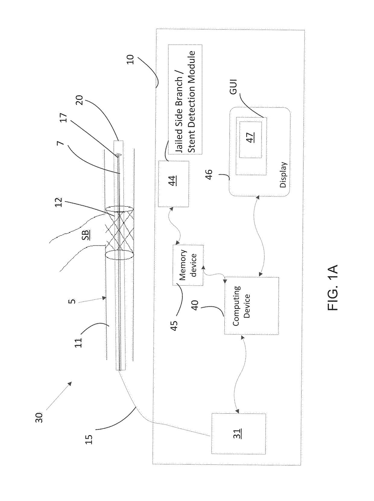
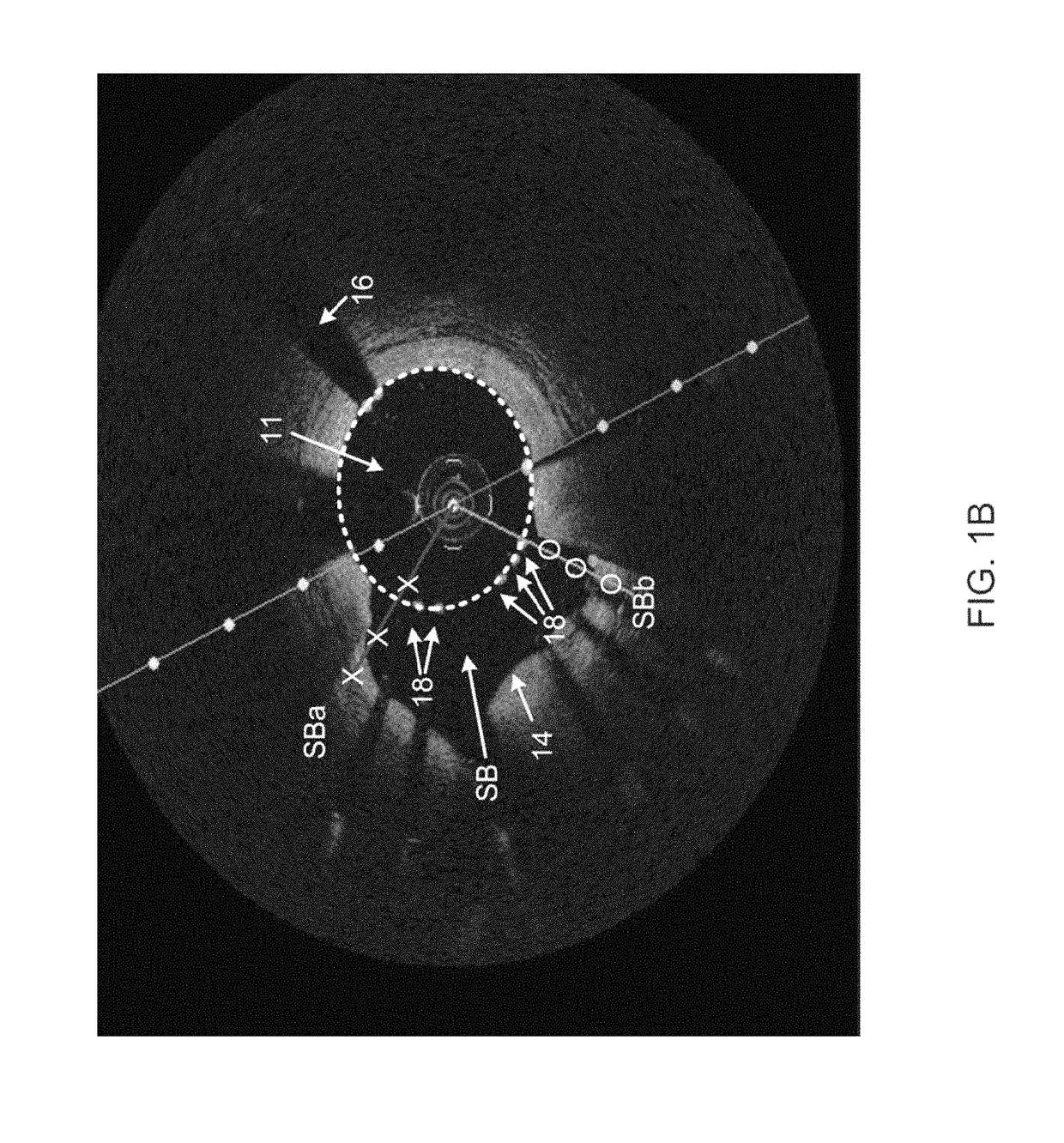
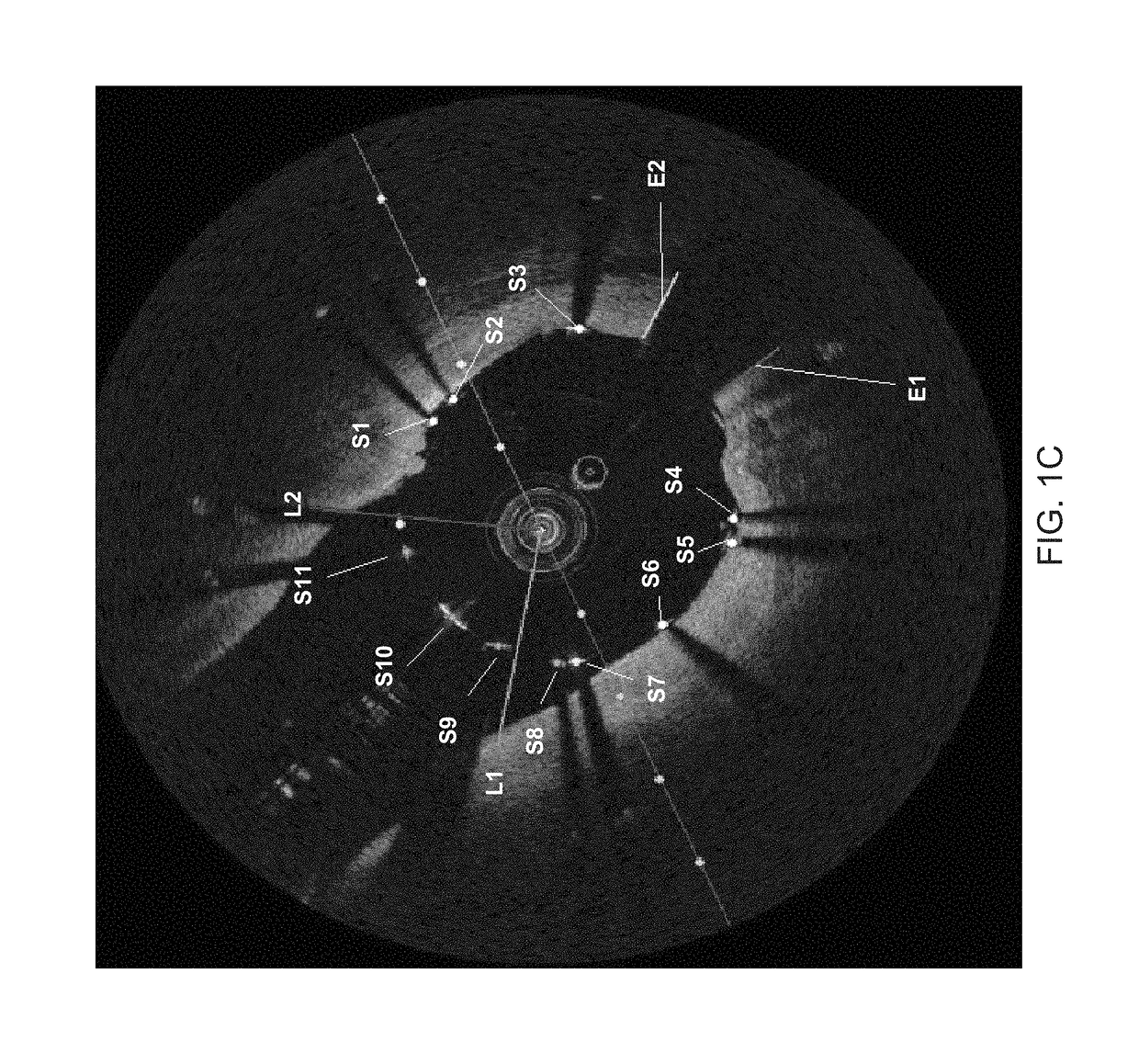
[0027]The systems and methods disclosed herein describe detecting and analyzing features of an artery using intravascular data including scan lines and images generated using scan lines or other data obtained with regard to the artery. In one embodiment, the intravascular data is analyzed and transformed to detect metal stent struts that block, cage, or otherwise “jail” a side branch of an artery. The intravascular data can include, for example, optical coherence tomography (OCT) or intravascular ultrasound (IVUS) data or other images of a blood vessel of interest. The intravascular data can be analyzed to identify sparse intensity peaks along each scan line—i.e., peaks that are surrounded by dark regions corresponding to a side branch which appears as a large shadow in most cases. In many cases a side branch manifests as an opening of the tissue region in the 2-D cross sectional view. As a consequence of this, there will be no shadows cast by the struts which jail the side branch.

[...


PUM


Abstract

In part, the disclosure relates to methods of stent strut detection relative to a side branch region using intravascular data. In one embodiment, detecting stent struts relative to jailed side branches is performed using a scan line-based peak analysis. In one embodiment, false positive determinations relating to stent struts are analyzed using a model strut.

Description

CROSS-REFERENCE TO RELATED APPLICATIONS[0001]This application claims priority to U.S. Provisional Patent Application No. 62 / 257,185 filed on Nov. 18, 2015, the disclosure of which is herein incorporated by reference in their entirety.FIELD[0002]The invention relates to systems and methods for stent detection.BACKGROUND[0003]Interventional cardiologists incorporate a variety of diagnostic tools during catheterization procedures in order to plan, guide, and assess therapies. Fluoroscopy is generally used to perform angiographic imaging of blood vessels. In turn, such blood vessel imaging is used by physicians to diagnose, locate and treat blood vessel disease during interventions such as bypass surgery or stent placement. Intravascular imaging technologies such as optical coherence tomography (OCT) are also valuable tools that can be used in lieu of or in combination with fluoroscopy to obtain high-resolution data regarding the condition of the blood vessels for a given subject.[0004]...

Claims


Application Information

Patent Timeline
Patent Type & Authority Patents(United States)
IPC IPC(8): G06K9/00G06T7/00
CPCG06T7/0014G06T2207/30101G06T2207/10101G06T7/0012G06T2207/10068A61B5/0066
Inventor AMBWANI, SONAL
Owner LIGHTLAB IMAGING
Why Eureka
Try Eureka