Method for differentiation of pluripotent stem cells into cardiomyocytes
a technology of stem cells and cardiomyocytes, applied in the field of differentiation of pluripotent stem cells into cardiomyocytes, can solve the problems of not being able to effectively generate cell models for drug development, not meeting the criteria above, and established cell models only partially reflect pharmaceutical relevan
- Summary
- Abstract
- Description
- Claims
- Application Information
AI Technical Summary
Benefits of technology
Problems solved by technology
Method used
Image
Examples
example 1
iation of Cardiomyocytes from Human Embryonic Stem Cells (hESC) and Induced Pluripotent Stem Cells (iPSC)
[0119]Human embryonic stem cells (hESC) or induced pluripotent stem cells (iPSC) were cultured in 56 cm2 dishes coated with Matrigel (BD Bioscience, Cat. 354277) at 37° C. and 5% CO2 in 10 ml mTeSR1 medium (Stemcell Technologies, Cat. 05850).
[0120]Before starting the cardiomyocyte differentiation, the cells were passaged 3-4 times to ensure that the pluripotent stem cells showed stable growth and duplication times.
[0121]To propagate pluripotent stem cells by conserving their pluripotent state, hESC or iPSC were treated with the following: the cells were washed once with 10 ml PBS− / −, and afterwards incubated with 3 ml Accutase (Innovative Cell Technologies, Cat. AT-104) for 2-3 minutes at 37° C. and 5% CO2, to detach the cells.
[0122]The enzymatic reaction of Accutase was stopped with 7 ml mTeSR1 and followed by centrifugation of the cells for 3 minutes at 500×g.
[0123]The cells we...
example 2
acterization
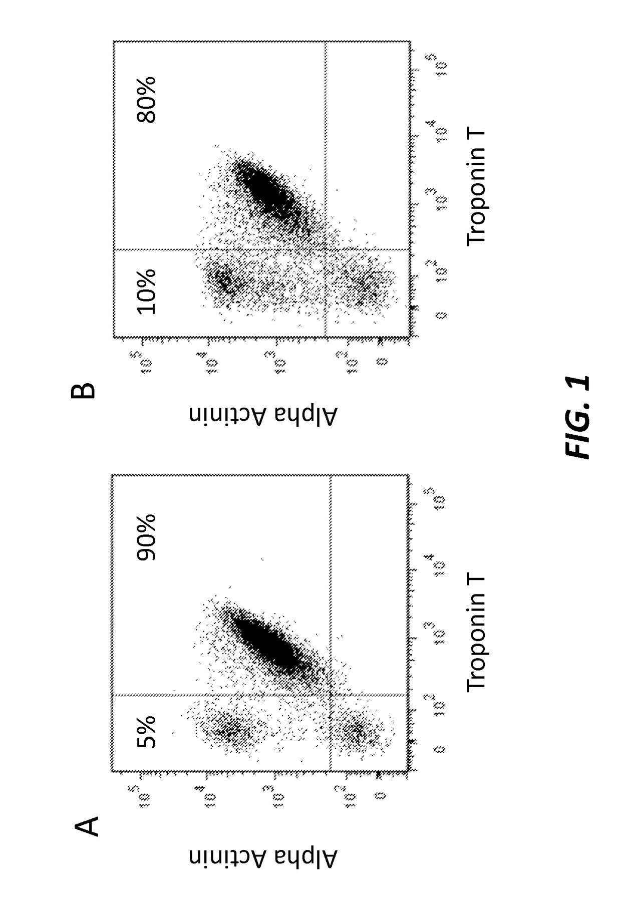
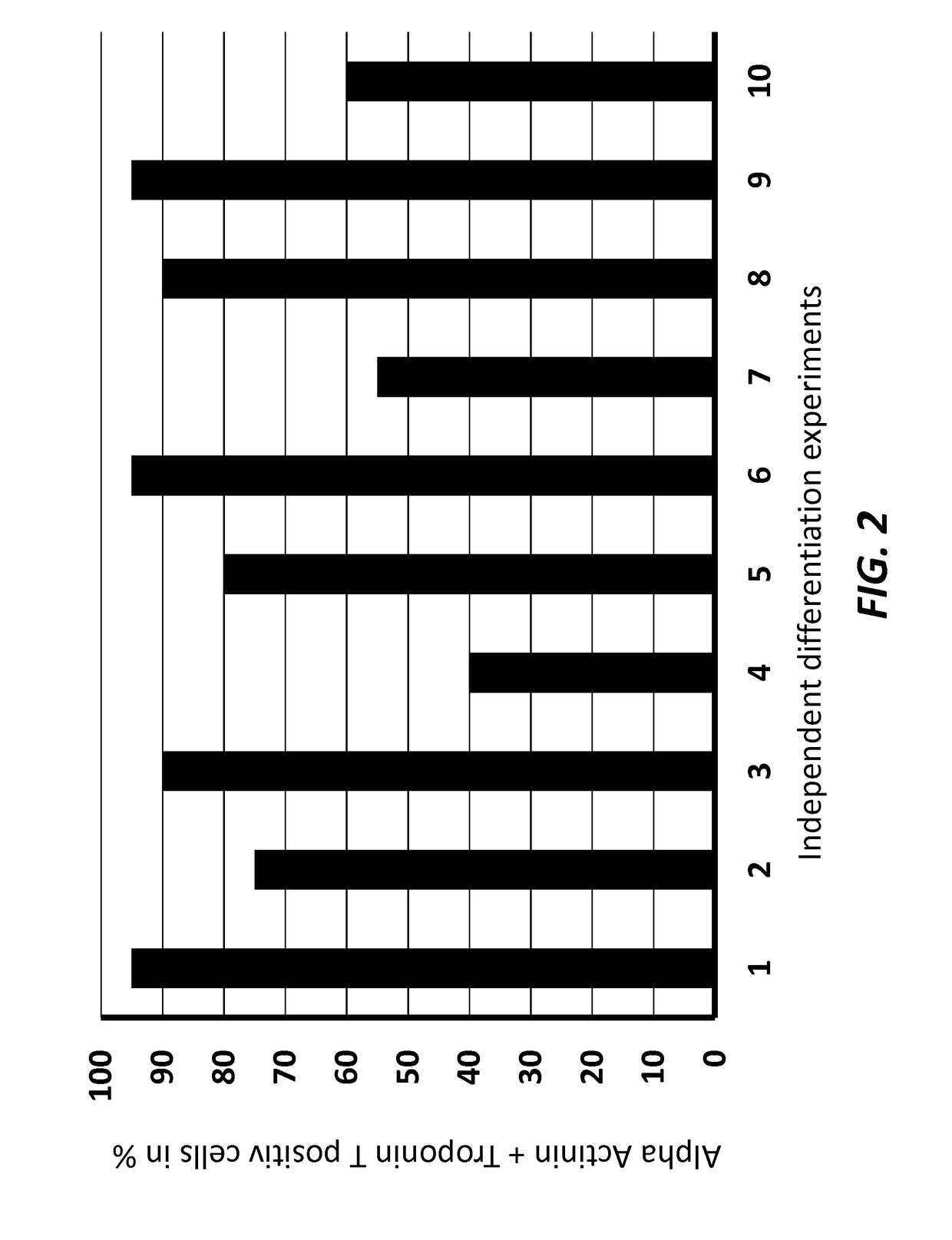
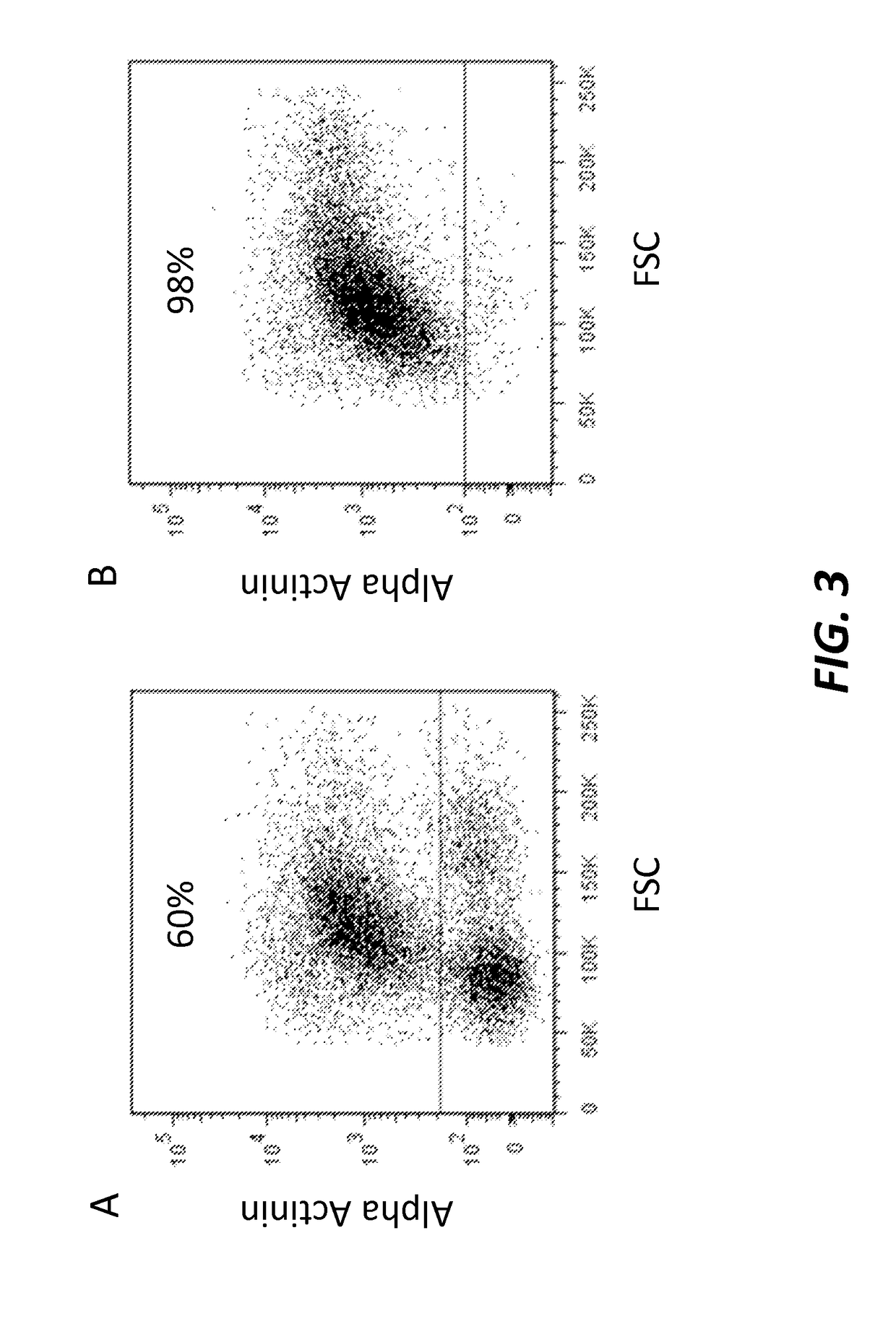
[0133]To test the efficiency of the differentiation process, the cardiomyocytes were characterized at differentiation day 14 by cell immunohistochemistry and Fluorescence Activated Cell Sorting (FACS) using antigens specific to cardiomyocytes.
[0134]Fluorescence Activated Cell Sorting (FACS) Analysis
[0135]Cells were washed with 180 μl / cm2 PBS− / − and dissociated 5-10 minutes with 100 μl / cm2 0.05% 1× Trypsin / EDTA (Gibco by Life Technologies, Cat. 25300) at 37° C. and 5% CO2.
[0136]If necessary, the cells were gently scraped from the cultivation vessel, pipetted up and down, and subsequently incubated 5-10 minutes at 37° C. and 5% CO2.
[0137]Afterwards threefold expansion medium and 10% fetal bovine serum (FBS) was added.
[0138]Then, the cells were filtered through 100 μm cell strainer and counted.
[0139]For analysis, 1×10⁶ cells in suspension were transferred into 1.5 ml tube. After 3 minutes centrifugation at 500×g, supernatant was removed and cells were fixed with 50 μl of In...
example 3
iation of Cardiomyocytes from Human Embryonic Stem Cells (hESC) and Induced Pluripotent Stem Cells (iPSC) Using Different CP21 Concentrations
[0143]The protocol as described above was repeated with different CP21 concentrations. The results are shown in Table 1 below: (−) No cardiomyocytes obtained, (+)—(+++): Amount of cardiomyocytes obtained.
[0144]
TABLE 1Cp21 conc. in μM00.3123510Experiment I−−++++++−Experiment II−−+++++−−Experiment III−−+++++−−
PUM
| Property | Measurement | Unit |
|---|---|---|
| time | aaaaa | aaaaa |
| density | aaaaa | aaaaa |
| time | aaaaa | aaaaa |
Abstract
Description
Claims
Application Information
 - R&D
- Intellectual Property
- Life Sciences
- Materials
- Tech Scout
- Unparalleled Data Quality
- Higher Quality Content
- 60% Fewer Hallucinations
Browse by: Latest US Patents, China's latest patents, Technical Efficacy Thesaurus, Application Domain, Technology Topic, Popular Technical Reports.
© 2025 PatSnap. All rights reserved.Legal|Privacy policy|Modern Slavery Act Transparency Statement|Sitemap|About US| Contact US: help@patsnap.com


