Method and wearable apparatus for disease diagnosis

a wearable apparatus and disease technology, applied in the field of image processing methods and equipment, can solve the problems of inability to immediately diagnose disease clinically, inability to observe with the naked eye, and inability to discover subtle variations

Active Publication Date: 2019-08-27
NAT TAIWAN UNIV
View PDF7 Cites 1 Cited by
  • Summary
  • Abstract
  • Description
  • Claims
  • Application Information

AI Technical Summary

Benefits of technology

[0007]The invention provides a method and a wearable apparatus for disease diagnosis which magnifies subtle variations on the surface of the human body and displays the magnified subtle variations on the wearable apparatus worn by a user, thereby realizing clinical disease diagnosis.
[0018]In view of the above, the wearable apparatus and the method for disease diagnosis of the present invention are directed to the images captured by the wearable apparatus. The images are split into various frequency bands according to the spatial frequency distribution thereof. After the images undergo time domain processing with respect to the frequency bands, a specific frequency range is then selected for analysis, thereby magnifying the subtle variations in the images. The magnified images may further be overlaid with the current image of the patient who is observed by medical personnel through the wearable apparatus. As a result, symptoms of the disease can be emphasized, and the clinical disease diagnosis can be achieved.

Problems solved by technology

This technology uncovers subtle variations that are undetectable or difficult to observe with the naked eye.
However, the scope of application has not been extended to instant disease diagnosis clinically yet.

Method used


Image

  • Method and wearable apparatus for disease diagnosis
  • Method and wearable apparatus for disease diagnosis
  • Method and wearable apparatus for disease diagnosis

Examples


Embodiment Construction

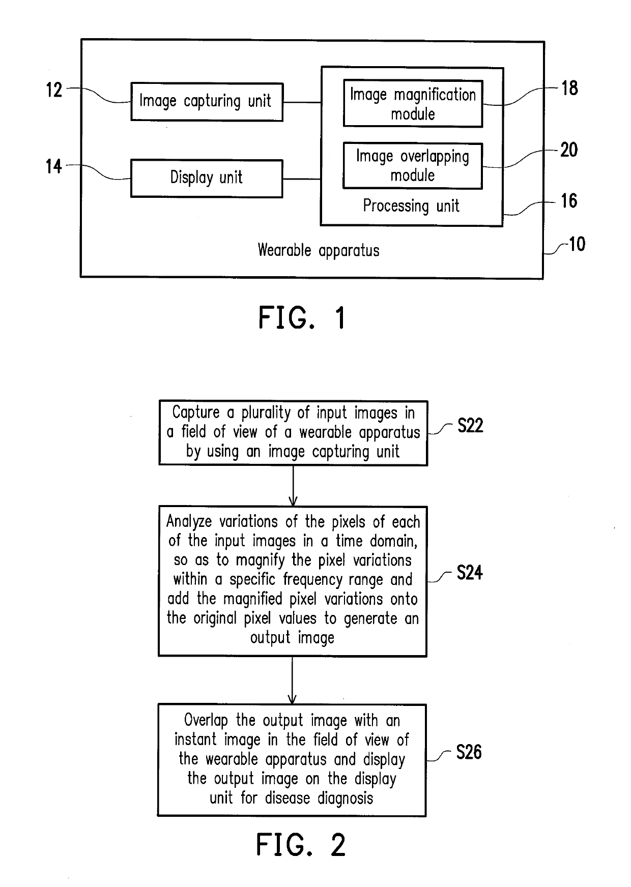
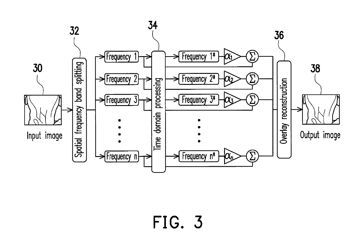
[0031]The invention combines image processing technology in a space domain and in a time domain, in which a subtle change in magnitude of an object captured by a wearable apparatus is brought into a phase observable by the naked eye via image processing and displayed on the wearable apparatus. When displaying a magnified image, the invention further performs feature analysis and comparison on the image and a current image captured by the wearable apparatus so as to overlap the image with an actual image observed by medical personnel through the wearable apparatus. By displaying the magnified symptom image on an actual object, medical personnel wearing the wearable apparatus may observe patient's symptoms immediately, thereby realizing clinical disease diagnosis.

[0032]FIG. 1 is a block diagram illustrating a wearable apparatus 10 according to an embodiment of the present invention. Referring to FIG. 1, the wearable apparatus 10 of the present embodiment is, for example, a device wear...


PUM


Abstract

A method and a wearable apparatus for disease diagnosis are provided. The method is applied to the wearable apparatus with an image capturing unit and a display unit. In this method, a plurality of input images in a field of view of the wearable apparatus are captured by using the image capturing unit, wherein each of the input images contains an array of pixels. The variations of the pixel values in a time domain are analyzed. The pixel variations within a specific frequency range are magnified and the magnified pixel variations are added onto the original ones to generate an output image. The output image is overlapped with a current image in the field of view of the wearable apparatus and displayed on the display unit.

Description

CROSS-REFERENCE TO RELATED APPLICATION[0001]This application claims the priority benefit of Taiwan application serial no. 103131894, filed on Sep. 16, 2014. The entirety of the above-mentioned patent application is hereby incorporated by reference herein and made a part of this specification.BACKGROUND OF THE INVENTION[0002]1. Field of the Invention[0003]The invention is related to an image processing method and apparatus, and more particularly, to a method and a wearable apparatus for disease diagnosis.[0004]2. Description of Related Art[0005]In recent years, new image processing technology that processes images by using computer algorithms to analyze and process variations of the images in a time domain has been introduced. This technology uncovers subtle variations that are undetectable or difficult to observe with the naked eye. Low amplitude movements that are unrecognizable by the naked eye, or even color changes due to blood flowing through the face, may all be brought into a...

Claims


Application Information

Patent Timeline
Patent Type & Authority Patents(United States)
IPC IPC(8): G06T7/00G06T7/33H04N5/262A61B3/00A61B3/14H04N5/265H04N5/232H04N5/225
CPCH04N5/2621G06T2207/10016G06T2207/10024G06T2207/20016G06T2207/30076G06T7/33G06T7/0016H04N5/265H04N23/51H04N23/635A61B3/14A61B3/0041
Inventor HSIAO, HAO-MINGKAO, HSIEN-LILEE, KUANG-HUEILI, DIAN-RU
Owner NAT TAIWAN UNIV
Why Eureka
Try Eureka