Image processing apparatus, image processing method, and image processing program

a technology of image processing and image processing method, which is applied in the direction of image enhancement, instruments, and recognition of medical/anatomical patterns, etc., can solve the problems of mammary glands, more likely to be hidden objects, and difficult to see in the case of mammary glands

Active Publication Date: 2021-03-23
FUJIFILM CORP
View PDF14 Cites 0 Cited by
  • Summary
  • Abstract
  • Description
  • Claims
  • Application Information

AI Technical Summary

Benefits of technology

[0028]According to the present disclosure, it is possible to detect a region in which an object of interest needs to be carefully observed in a radiographic image of the breast.

Problems solved by technology

However, even though the mammary gland content rate of the entire breast is the same in a case in which the mammary glands are concentrated in some regions of the breast and in a case in which the mammary glands are scattered in the entire breast, the object of interest is more likely to be hidden and is more difficult to see in the case in which the mammary glands are concentrated.

Method used


Image

  • Image processing apparatus, image processing method, and image processing program
  • Image processing apparatus, image processing method, and image processing program
  • Image processing apparatus, image processing method, and image processing program

Examples


first embodiment

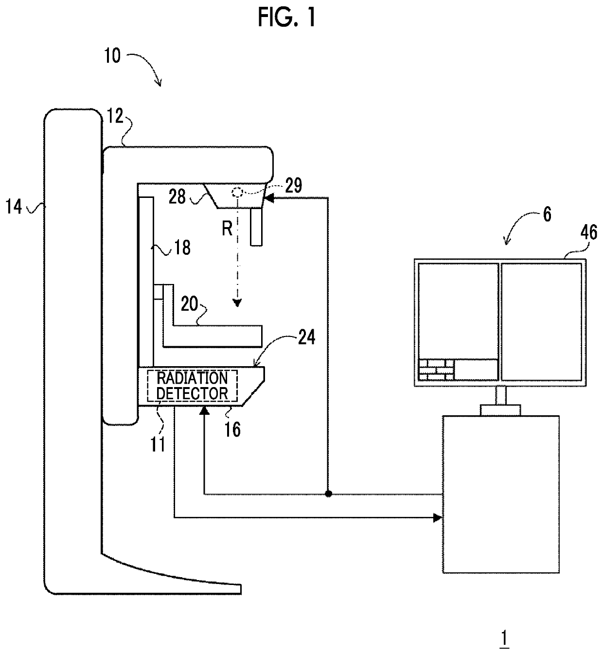
[0047]First, an example of the overall configuration of a radiography system according to this embodiment will be described. FIG. 1 is a configuration diagram illustrating an example of the overall configuration of a radiography system 1 according to this embodiment.

[0048]The radiography system 1 according to this embodiment has a function of capturing radiographic images in response to an operation of a user, such as a doctor or a radiology technician, on the basis of a command (imaging order) input from an external system (for example, a radiology information system (RIS)) through a console 6.

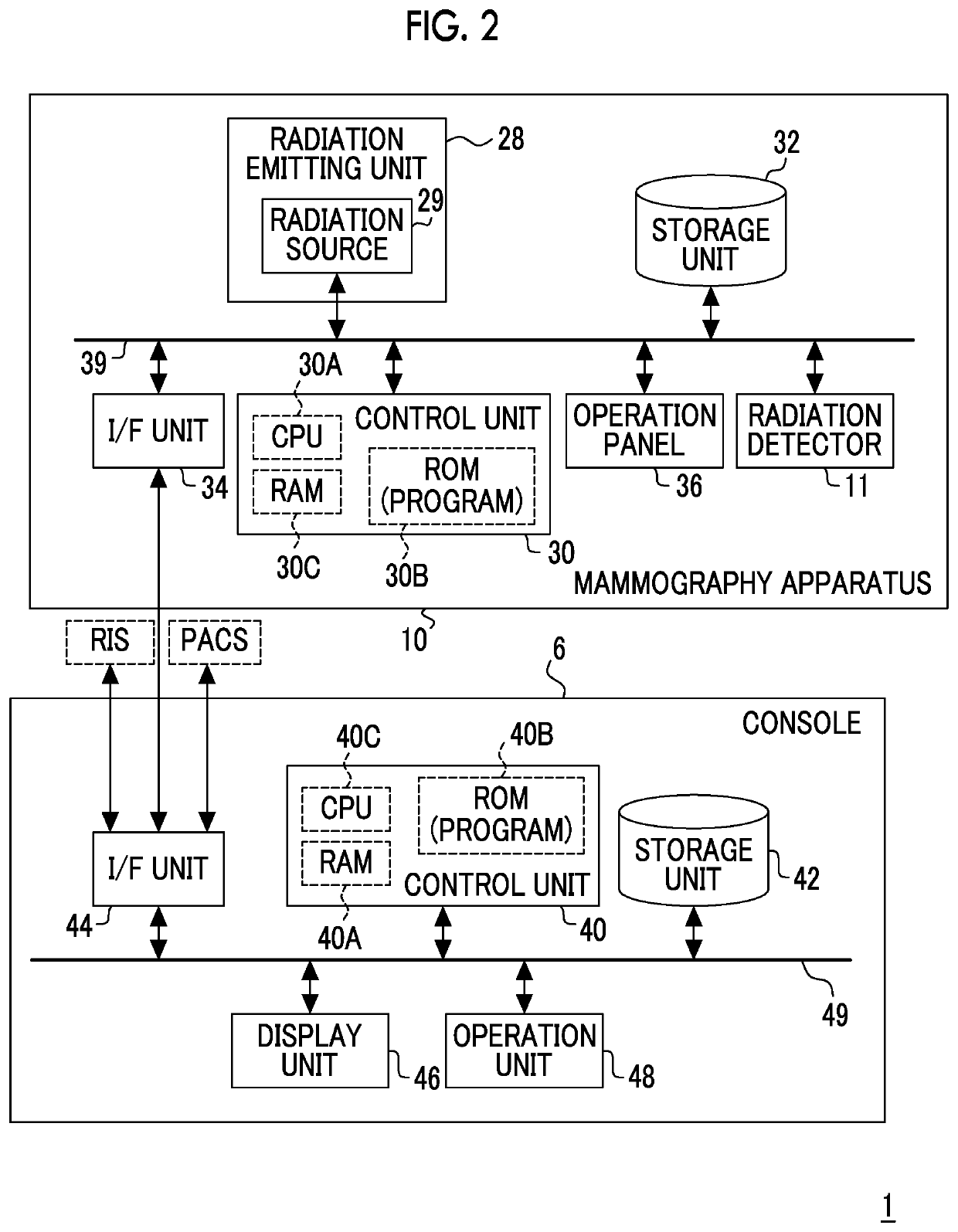
[0049]As illustrated in FIG. 1, the radiography system 1 according to this embodiment includes the console 6 and a mammography apparatus 10. FIG. 2 is a block diagram illustrating an example of the configuration of the console 6 and the mammography apparatus 10 according to this embodiment.

[0050]The console 6 according to this embodiment has a function of controlling the mammography apparatus...

second embodiment

[0106]Next, a second embodiment will be described in detail. In this embodiment, the same configurations and operations as those in the first embodiment are denoted by the same reference numerals and the detailed description thereof will not be repeated.

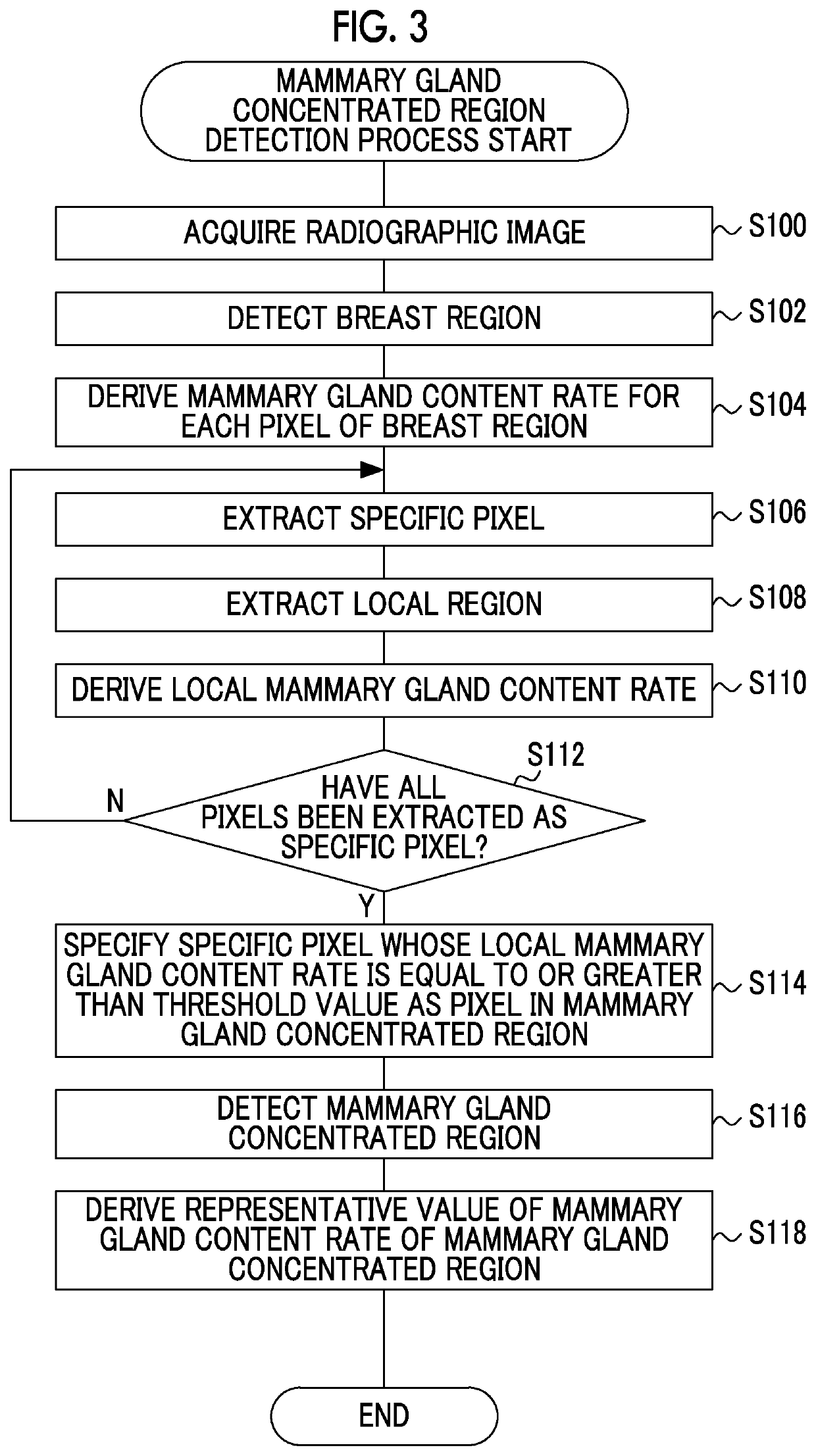
[0107]Since the configurations of a radiography system 1, a console 6, and a mammography apparatus 10 are the same as those in the first embodiment, the description thereof will not be repeated. In this embodiment, since a mammary gland concentrated region detection process performed by the control unit 40 of the console 6 is partially different from the mammary gland concentrated region detection process (see FIG. 3) in the first embodiment, different processes will be described.

[0108]FIG. 11 is a flowchart illustrating an example of the flow of the mammary gland concentrated region detection process in the console 6 according to this embodiment. In a case in which the mammary gland concentrated region detection process illustrated ...

third embodiment

[0116]Next, a third embodiment will be described in detail. In this embodiment, the same configurations and operations as those in the first embodiment are denoted by the same reference numerals and the detailed description thereof will not be repeated.

[0117]Since the configurations of a radiography system 1 and a console 6 are the same as those in the first embodiment, the description thereof will not be repeated. In this embodiment, the configuration of a mammography apparatus 10 is partially different from that in the first embodiment. The mammography apparatus 10 according to this embodiment has a so-called tomosynthesis imaging function which irradiates the breast with the radiation R emitted from the radiation source 29 at each of a plurality of irradiation angles to capture a plurality of projection images.

[0118]FIG. 12 is a block diagram illustrating an example of the configuration of the console 6 and the mammography apparatus 10 according to this embodiment. As illustrated...


PUM


Abstract

In a console, a control unit functions as an acquisition unit that acquires a radiographic image of a breast, a derivation unit that derives a mammary gland content rate for each pixel of a breast region in the radiographic image, and a detection unit that detects a mammary gland concentrated region in which mammary glands are concentrated on the basis of a result of specifying whether a specific pixel which is each pixel of the breast region is a pixel included in the mammary gland concentrated region of the breast region on the basis of the mammary gland content rate of the specific pixel and a mammary gland content rate of a pixel in a local region around the specific pixel.

Description

CROSS-REFERENCE TO RELATED APPLICATIONS[0001]The present application claims priority under 35 U.S.C. § 119 to Japanese Patent Application No. 2018-068965, filed on Mar. 30, 2018. Each of the above application(s) is hereby expressly incorporated by reference, in its entirety, into the present application.BACKGROUND OF THE INVENTION1. Field of the Invention[0002]The present disclosure relates to an image processing apparatus, an image processing method, and a non-transitory computer readable recording medium storing an image processing program.2. Description of the Related Art[0003]In general, an object of interest, such as a breast cancer, in the breast has been diagnosed on the basis of a radiographic image of the breast of a subject captured by a so-called mammography apparatus. However, in the case of the breast with high mammary gland density, an image of the object of interest in the radiographic image may be hidden by the mammary glands and may be difficult to see. Therefore, a...

Claims


Application Information

Patent Timeline
Patent Type & Authority Patents(United States)
IPC IPC(8): G06K9/00G06T7/00G06K9/32A61B5/00G06V10/25
CPCG06T7/0012A61B5/0091A61B5/743G06K9/3233G06T2207/10081G06T2207/30068G06T2207/10116A61B6/502A61B6/5217A61B6/12A61B6/463A61B6/0414A61B6/025A61B6/563G06V10/25G06V2201/03
Inventor MORITA, JUNYA
Owner FUJIFILM CORP
Why Eureka
Try Eureka