
Methods, apparatuses, and systems for creating 3-dimensional representations exhibiting geometric and surface characteristics of brain lesions

a technology of geometric and surface characteristics and brain lesions, applied in the field of brain lesions geometric and surface characteristics, can solve the problems of heterogeneity of lesions, difficult to accurately assess the size, volume, and surface area of lesion, and the inability to effectively apply the existing dissemination criteria. , to achieve the effect of redefining the shape and surface features, accurate assessment of lesion size, volume and surface area

Active Publication Date: 2021-08-17
BOARD OF RGT THE UNIV OF TEXAS SYST
View PDF31 Cites 4 Cited by
  • Summary
  • Abstract
  • Description
  • Claims
  • Application Information

AI Technical Summary

Benefits of technology

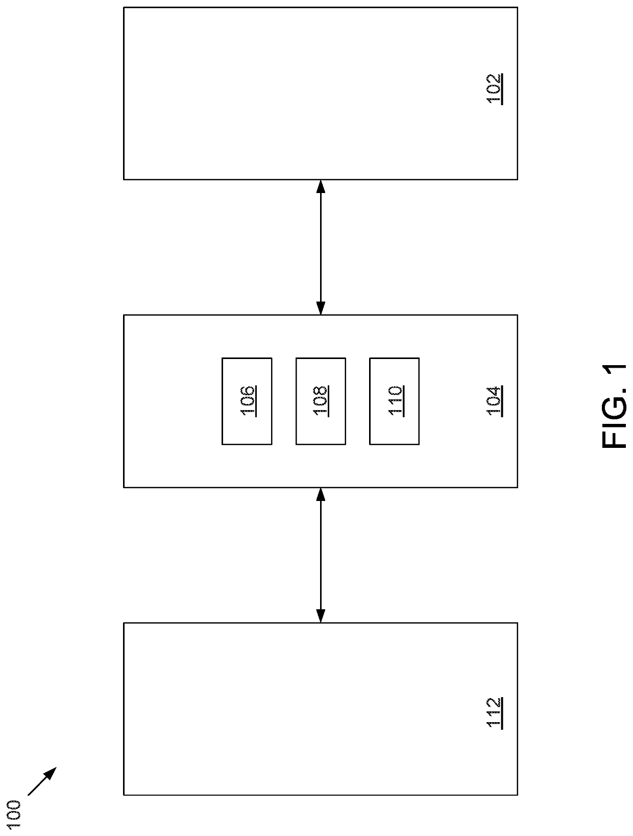
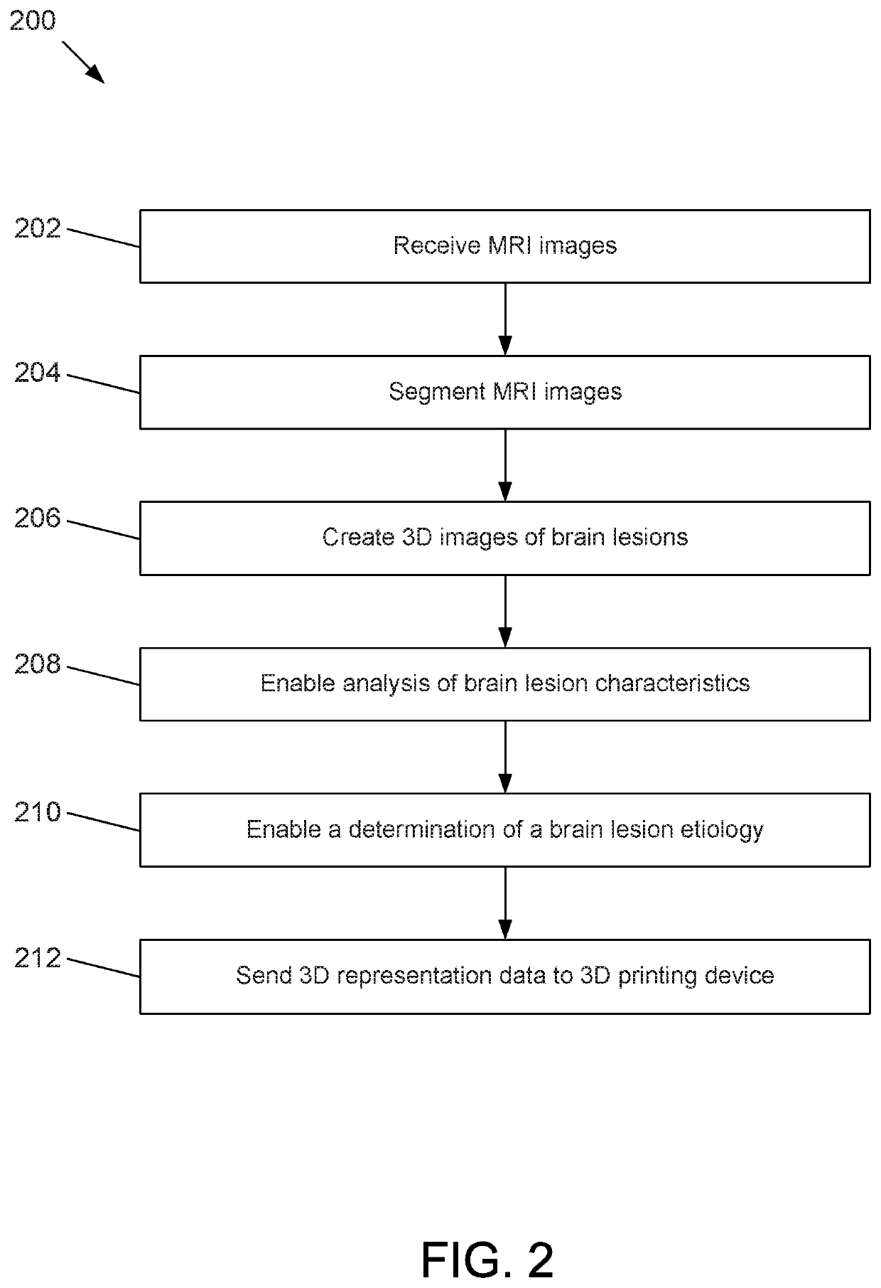
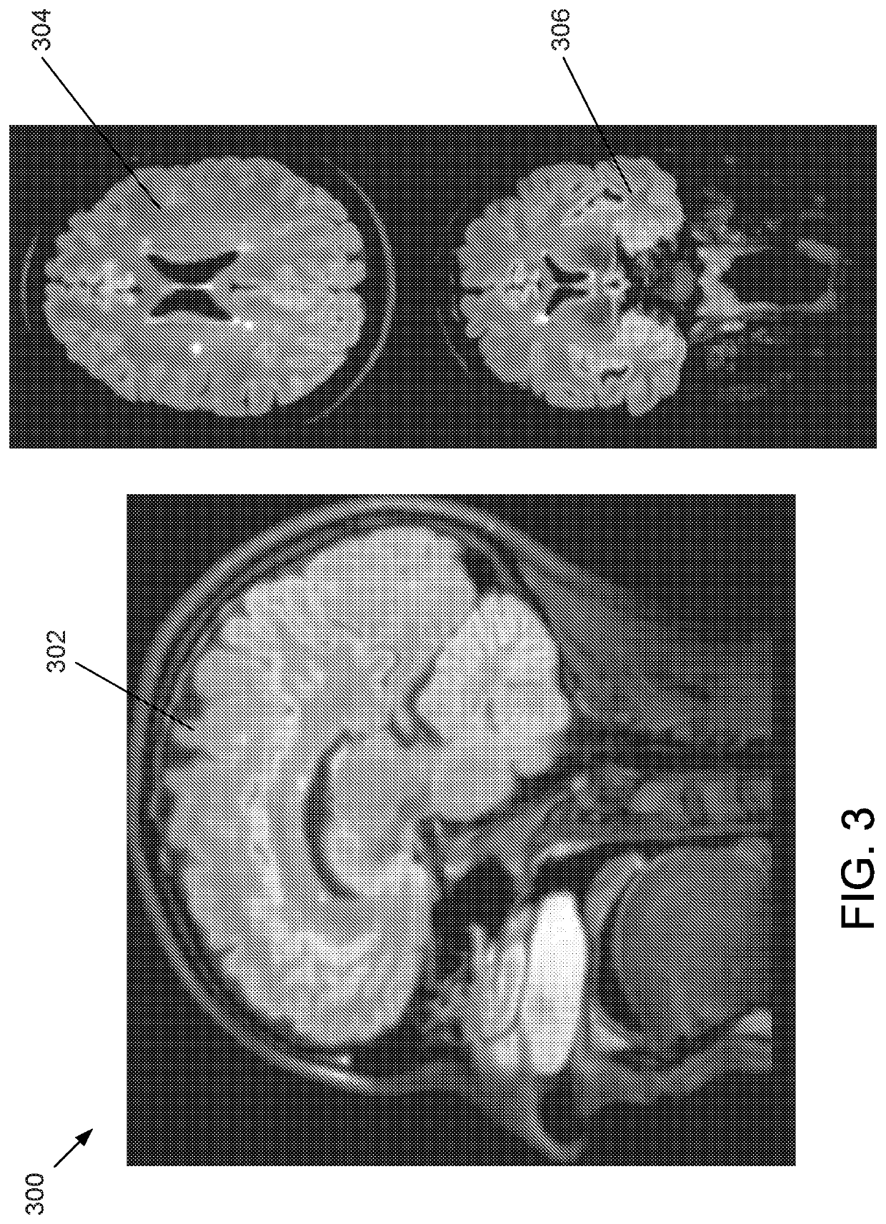
[0005]This disclosure includes embodiments that allow for the depiction of MRI brain lesions in true 3-dimensional (3D) form. This approach may isolate lesions resulting from patients with multiple sclerosis and those with non-specific white matter disease changes due to normal aging, migraine headaches, high blood pressure, smoking, and other vascular risk factors. By isolating and evaluating brain lesions in actual 3D form, in comparison to conventional 2D approaches that provide slice-by-slice renditions of 3D shapes, the disclosed embodiments may enable a better understanding of both lesion shapes and surface features in vivo and new descriptive characteristics may be generated based on the findings between groups. The unique approach of the disclosed embodiments represents a scientific effort aimed at redefining the shapes and surface features of MS lesions compared with those resulting from one of the most common disease states responsible for white matter injury within the brain—non-specific white matter (NSWM) disease. Beyond geometric shape and surface characteristics, the disclosed embodiments may also provide a more accurate assessment of lesion sizes, volumes, and surface areas when compared to existing technology.

Problems solved by technology

The effective application of the existing dissemination in space criteria may be hindered by the highly sensitive nature of magnetic resonance imaging (MRI) technology, the heterogeneity of lesions resulting from a variety of medical conditions, concomitant radiological features resulting from age-related changes and disease, and the lack of additional radiological characteristics beyond 2-dimensional (2D) descriptions.
However, this technique has been limited by the lack of appreciation of the central vessel in all orthogonal planes of view and the abundance of vessels intersecting lesions within the supratentorial region.

Method used


Image

  • Methods, apparatuses, and systems for creating 3-dimensional representations exhibiting geometric and surface characteristics of brain lesions
  • Methods, apparatuses, and systems for creating 3-dimensional representations exhibiting geometric and surface characteristics of brain lesions
  • Methods, apparatuses, and systems for creating 3-dimensional representations exhibiting geometric and surface characteristics of brain lesions

Examples


example experimental

[0056 results are depicted in FIGS. 6-9. In the embodiment shown in FIG. 6, results 600 may include one or more 2D MRI images 602, one or more 3D MRI images 604, one or more 3D brain lesion images 606, and one or more 3D physical brain lesion representations 608. In the embodiment shown, one or more 2D MRI images 602 are axial MRI FLAIR images of the brain, one or more 3D MRI images 604 are MIP images, one or more 3D brain lesion images 606 are orthographic images, and one or more 3D physical brain lesion representations 608 are 3D printed regions of interest from four unique patients. In the embodiment shown, images A-D exhibit asymmetric primary lesion characteristics, images E-H exhibit symmetric primary lesion characteristics, images I-L exhibit complex surface morphology primary lesion characteristics, and images M-P exhibit simple surface morphology primary lesion characteristics.

[0057]In the embodiment shown in FIG. 7, results 700 may include one or more 2D MRI images 702, on...


PUM


Abstract

Methods, apparatuses, systems, and implementations for creating 3-dimensional (3D) representations exhibiting geometric and surface characteristics of brain lesions are disclosed. 2D and / or 3D MRI images of the brain may be acquired. Brain lesions and other abnormalities may be identified and isolated with each lesion serving as a region of interest (ROI). Saved ROI may be converted into stereolithography format, maximum intensity projection (MIP) images, and / or orthographic projection images. Data corresponding to these resulting 3D brain lesion images may be used to create 3D printed models of the isolated brain lesions using 3D printing technology. Analysis of the 3D brain lesion images and the 3D printed brain lesion models may enable a more efficient and accurate way of determining brain lesion etiologies.

Description

CROSS-REFERENCE TO RELATED APPLICATION[0001]This application is a national phase application under 35 U.S.C. § 371 of International Application No. PCT / US2017 / 040229, filed Jun. 30, 2017 which claims priority to U.S. Provisional Patent Application 62 / 357,477 filed Jul. 1, 2016, the content of each is incorporated herein by reference in its entirety.BACKGROUND1. Field of the Invention[0002]This disclosure relates generally to methods, apparatuses, and systems for creating 3-dimensional (3D) representations exhibiting geometric and surface characteristics of brain lesions.2. Description of Related Art[0003]The diagnosis of multiple sclerosis (MS) requires the fulfillment of both clinical and radiological criteria. This may include a determination of key radiological tenets such as a requisite number of lesions having a specific character (i.e. size, shape, and morphology) and spatial distribution patterns with involvement of periventricular, juxtacortical, infratentorial, and spinal c...

Claims


Application Information

Patent Timeline
Patent Type & Authority Patents(United States)
IPC IPC(8): G06K9/62G06K9/34G06T7/11G06T15/08G06T17/00G16H30/40A61B5/055G06V10/26G06V10/75
CPCG06K9/6201G06K9/34G06T7/11G06T15/08G06T17/00A61B5/055G06K2209/051G06T2207/10088G06T2207/30016G06T2207/30096G06T2210/41G16H30/40G06V10/26G06V2201/031G06V10/75G06F18/22
Inventor OKUDA, DARIN TNEWTON, BRAEDEN D
Owner BOARD OF RGT THE UNIV OF TEXAS SYST
Learn More