Detection of nuclease edited sequences in automated modules and instruments

a technology of automated modules and sequences, applied in biochemistry apparatus and processes, laboratory glassware, tissue/virus culture apparatus, etc., can solve the problems of skewed representations of different edits in the population, high editing efficiency in cell populations, and difficulty in identifying edited cells in the background of unedited cells, so as to improve the observed editing efficiency, and double the observed editing efficiency

Active Publication Date: 2021-10-12
INSCRIPTA INC
View PDF114 Cites 2 Cited by
  • Summary
  • Abstract
  • Description
  • Claims
  • Application Information

AI Technical Summary

Benefits of technology

[0007]The present disclosure provides methods, modules and instruments for automated high-throughput and extremely sensitive enrichment and selection of cells edited by a nucleic acid-guided nuclease. Some embodiments of the methods take advantage of isolation or substantial isolation, e.g., separating individual cells, providing conditions for editing, and growing the individual cells into clonal colonies. Isolation overcomes growth bias from unedited cells, growth effects from differential editing rates, and growth bias resulting from fitness effects of different edits. Indeed, it has been determined that removing growth rate bias via isolation and growing colonies from the isolated cells to saturation or terminal colony size improves the observed editing efficiency by up to 4× or more over conventional methods. Other embodiments of the methods utilize “cherry picking” or direct selection of edited colonies, determined by the differential growth rate of edited and unedited cell colonies. Cherry picking colonies using the methods described herein more than doubles the observed editing efficiency as the result of isolation. Thus, the combination of isolation and cherry picking improves observed editing efficiency by up to 98% over conventional methods.
[0008]The present disclosure provides instruments, modules and methods to enable automated high-throughput and extremely sensitive screening to identify edited cells in populations of cells that have been subjected to nucleic acid-guided nuclease editing. The instruments, modules, and methods take advantage of isolation or substantial isolation, where the term “isolation” in this context refers to the process of separating cells and growing them into clonally-isolated formats. The term “substantial isolation” refers to the process of separating cells in a population of cells into “groups” of 2 to 100, or 2 to 50, and preferably 2 to 10 cells. Isolation (or substantial isolation), followed by an initial period of growth (e.g., incubation), editing, and growth normalization leads to enrichment of edited cells. Further, certain of the instruments, modules, and methods described herein facilitate “cherry picking” of edited cell colonies, allowing for direct selection of edited cells.
[0009]Isolation or substantial isolation assists in overcoming the growth bias from unedited cells that occurs under competitive growth regimes such as in bulk liquid culture. Indeed, it has been determined that removing growth rate bias via isolation or substantial isolation, incubation, editing and normalization improves the observed editing efficiency by up to 4× (from, e.g., 10% to 40% absolute efficiency at population scale) or more over conventional methods, and further that cherry-picking colonies using the methods described herein brings the observed editing efficiency up to 8× (from, e.g., 10% to 80% absolute efficiency at population scale) over conventional methods. In some embodiments—particularly in bacteria—the compositions and methods employ inducible guide RNA (gRNA) constructs leading to increased observed transformation efficiency and automation-friendly control over the timing and duration of the editing process.
[0010]One particularly facile module or device for isolation or substantial isolation is a solid wall device where cells are substantially isolated, grown in a clonal (or substantially clonal) format, edited, and either normalization or cherry picking is employed. The solid wall devices or modules and the uses thereof are described in detail herein. The instruments, modules and methods in some embodiments allow for normalization of edited and unedited cell colonies. Normalization refers to growing colonies of cells—whether edited or unedited—to terminal size; that is, growing the cells until the cells in the colonies enter senescence due to, e.g., nutrient exhaustion or constrained space for further growth. Since unedited cells grow more quickly, unedited cell colonies will reach terminal size (e.g., senescence) before edited cell colonies; however, the unedited cell colonies eventually “catch up” in size and senescence. Thus, normalization of cell colonies enriches for edited cells as edited cells get “equal billing” with unedited cells. Additionally, certain of the modules as described herein facilitate “cherry picking” of colonies. Cherry picking allows for direct selection of edited cells by taking advantage of edit-induced growth delay in edited colonies. Cherry picking can be performed by selecting slow-growing cell colonies, or cherry picking can be performed by eliminating faster-growing cell colonies by, e.g., irradiating the faster-growing cell colonies in the wells in which they are growing. Cherry picking colonies using the instruments, modules, and methods described herein may more than double the observed editing efficiency as the result of isolation or substantial isolation alone.

Problems solved by technology

Editing efficiencies in cell populations can be high; however, in pooled or multiplex formats there tends to be selective enrichment of cells that have not been edited due to the lack of the double-strand DNA breaks that occur during the editing process.
Double-strand DNA breaks dramatically negatively impact cell viability thereby leading to the enhanced survival of unedited cells and making it difficult to identify edited cells in the background of unedited cells.
In addition, cells with edits that confer growth advantages or disadvantages can lead to skewed representations for different edits in the population.

Method used


Image

  • Detection of nuclease edited sequences in automated modules and instruments
  • Detection of nuclease edited sequences in automated modules and instruments
  • Detection of nuclease edited sequences in automated modules and instruments

Examples


example 1

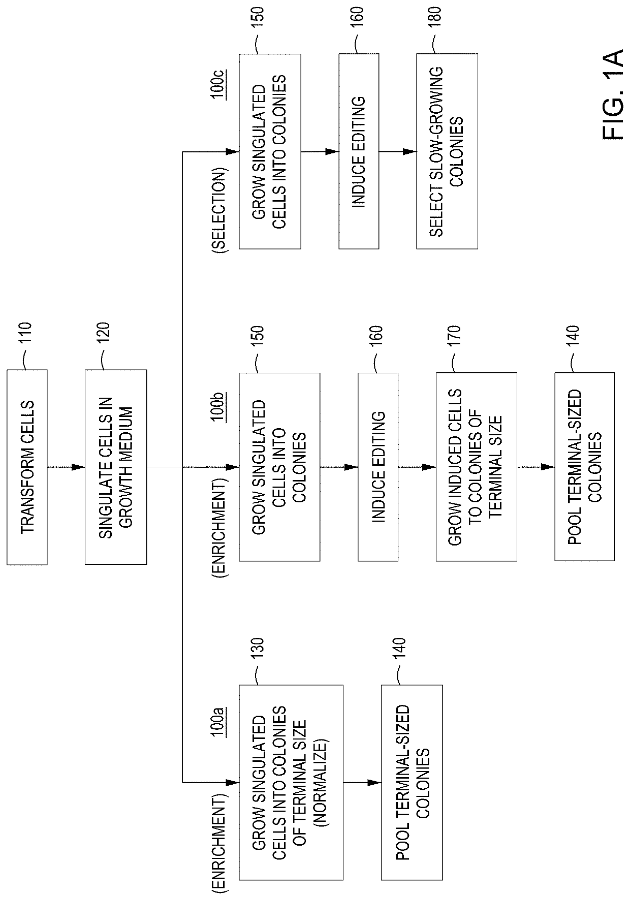
t of Editing Cells by Growth Lag Identification

[0210]Transformation:

[0211]100 ng of the editing vector cloned library or Gibson Assembly® reaction was transformed by electroporation into 100 μL competent EC1 cells containing the engine vector. The electroporator was set to 2400 V in 2 mm cuvette. Following transformation, the cells were allowed to recover for 3 hours in SOB medium. A 10-fold dilution series of recovered cells (in H2O) was spot plated and the resulting CFU counts / dilution ratios were used to calculate transformation efficiency.

[0212]Plating and Colony Arraying:

[0213]100 μL of the appropriate dilution was plated on LB medium+25 μg / mL chlor and +arabinose and grown at 30° C. for 6-8 hours. Alternatively, the cells may be grown in liquid culture in LB medium+25 μg / mL chlor at 30° C. for 6-8 hours. The temperature of the plate was adjusted to 42° C. and the plates were incubated for two hours. The temperature was then adjusted back to 30° C. and the cells were allowed to...

example 2

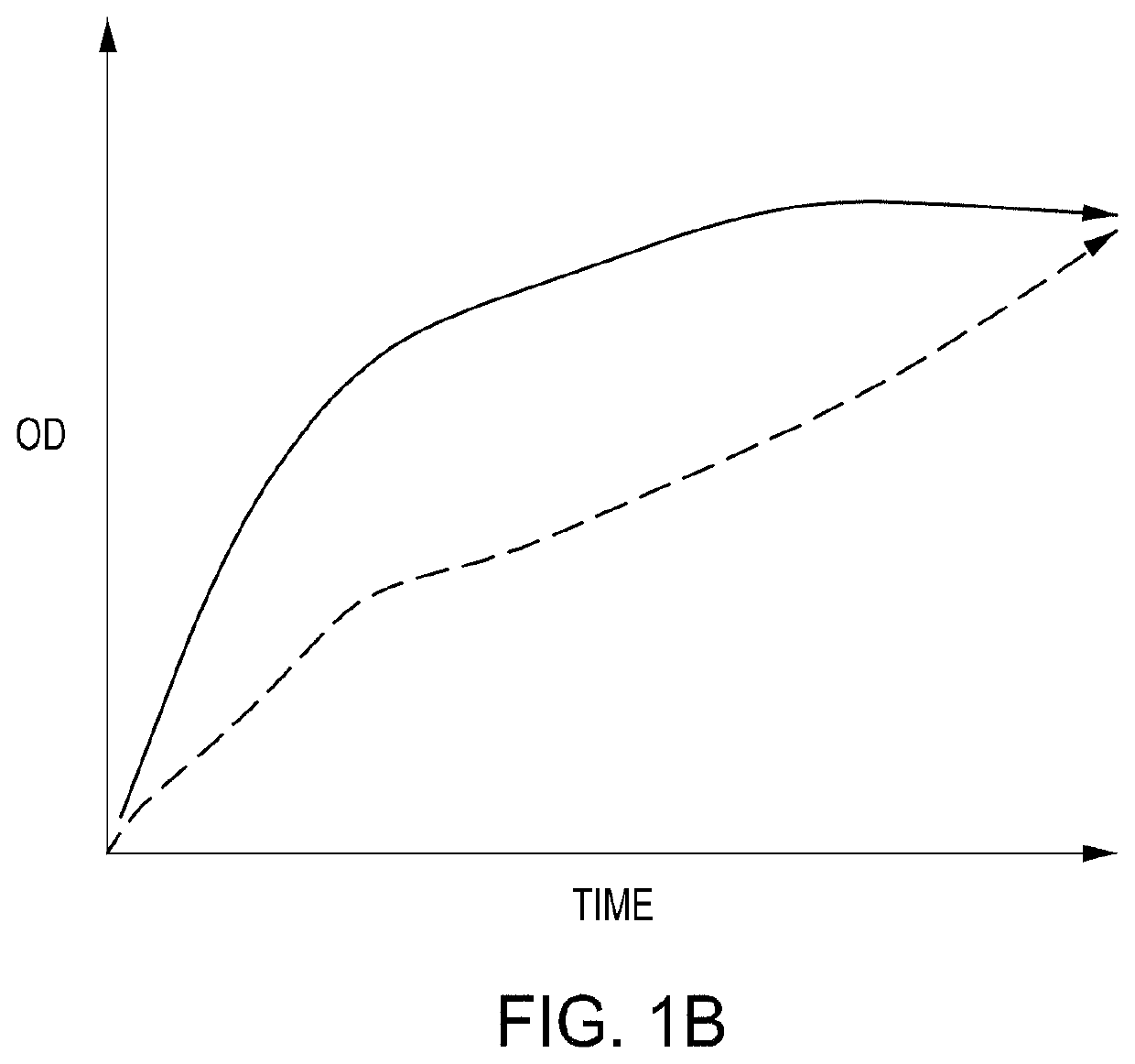
Editing by Optical Density

[0216]FIG. 2C is a depiction of the growth profiles of randomly-picked variants from a silent PAM mutation (SPM) library. This 500-member library targets regions located across the entire E. coli genome and integrated synonymous mutations that have no expected fitness effects. Colonies were picked from agar plates of uninduced transformed cells. Cells were picked from an agar plate and grown up in 200 μL LB+chlor / carb overnight in a 96-well microtiter plate format. 10 μL of the well content of the parent microtiter plate was then transferred to two replica daughter microtiter plates that received either no induction (top) or gRNA and nuclease induction (via the pL inducible promoter) for 1 hour at 42° C. (bottom). The well maps show the relative OD at 6 hours; the full growth curves are shown for reference. The replica wells represent growth observed from the same cassette design with or without gRNA induction. While the majority of the wells for the no-ind...

example 3

assette and Backbone Amplification and Assembly

[0217]Editing Cassette Preparation:

[0218]5 nM of oligonucleotides synthesized on a chip were amplified using Q5 polymerase in 50 μL volumes. The PCR conditions were 95° C. for 1 minute; 8 rounds of 95° C. for 30 seconds / 60° C. for 30 seconds / 72° C. for 2.5 minutes; with a final hold at 72° C. for 5 minutes. Following amplification, the PCR products were subjected to SPRI cleanup, where 30 μL SPRI mix was added to the 50 μL PCR reactions and incubated for 2 minutes. The tubes were subjected to a magnetic field for 2 minutes, the liquid was removed, and the beads were washed 2× with 80% ethanol, allowing 1 minute between washes. After the final wash, the beads were allowed to dry for 2 minutes, 50 μL 0.5×TE pH 8.0 was added to the tubes, and the beads were vortexed to mix. The slurry was incubated at room temperature for 2 minutes, then subjected to the magnetic field for 2 minutes. The eluate was removed and the DNA quantified.

[0219]Foll...


PUM

PropertyMeasurementUnit
volumeaaaaaaaaaa
volumeaaaaaaaaaa
volumeaaaaaaaaaa

Abstract

The present disclosure provides automated modules and instruments for improved detection of nuclease genome editing of live cells. The disclosure provides improved modules—including high throughput modules—for screening cells that have been subjected to editing and identifying and selecting cells that have been properly edited.

Description

RELATED APPLICATIONS[0001]This application claims priority to U.S. Provisional Application Nos. 62 / 718,449, filed 14 Aug. 2018; 62 / 769,805, filed 20 Nov. 2018; 62 / 735,365, filed 24 Sep. 2018; 62 / 781,112, filed 18 Dec. 2018; 62 / 779,119, filed 13 Dec. 2018; and 62 / 841,213, filed 30 Apr. 2019. This application is also related to U.S. application Ser. No. 16 / 399,988, filed 30 Apr. 2019; and Ser. No. 16 / 454,865, filed 26 Jun. 2019, all of which are incorporated herein in their entirety.FIELD OF THE INVENTION[0002]This invention relates to automated modules and instruments for screening, identifying, and selecting live cells that have nuclease-directed edits.BACKGROUND OF THE INVENTION[0003]In the following discussion certain articles and methods will be described for background and introductory purposes. Nothing contained herein is to be construed as an “admission” of prior art. Applicant expressly reserves the right to demonstrate, where appropriate, that the methods referenced herein d...

Claims


Application Information

Patent Timeline
Patent Type & Authority Patents(United States)
IPC IPC(8): C12M3/00C12N15/00B01L3/00C12M1/42C12N15/11C12N15/90C12M1/26
CPCC12M35/08C12M23/44C12M33/14C12N15/11C12N15/902C12N2310/20C12M35/02
Inventor GARST, ANDREWGRAIGE, MICHAELFOX, RICHARDSPINDLER, EILEENHIDDESSEN, AMYBELGRADER, PHILLIPMASQUELIER, DONCHABANSKY, BRUCE
Owner INSCRIPTA INC
Why Eureka
Try Eureka