X-ray imaging device

Active Publication Date: 2022-02-01
ADAPTIX
View PDF5 Cites 0 Cited by
  • Summary
  • Abstract
  • Description
  • Claims
  • Application Information

AI Technical Summary

Benefits of technology

[0021]A further benefit of the use of arrays of angled panels is the ability to better locate or determine the relative position of the source relative to the detector. In many clinical applications, for instance dental and in-bed imaging, the location of the detector is not well known as it is hidden (e.g. inside the mouth or behind the patient) and the relative position of the source

Problems solved by technology

It is also known that multiple x-ray tubes can be arranged to accomplish similar tasks and with greater speed and precision, but with added cost and complexity.
As a consequence of the source geometry, the configurations possible for x-ray imaging systems are limited and generally include 2D “planar”, 3D “CT” (computed tomography), and partially-3D “tomosynthesis” systems.
Many of these deficiencies come from an inability to remove the impact (attenuation) of overlying and underlying tissue.
CT generally addresses the deficiencies of planar imaging, however, at an order of magnitude increase in the cost of deployment, cost per use, dose to the patient, size, weight (and hence lack of portability), read time, etc.
In critical care or serious conditions, the dose burden and costs are generally justified, but in more routine imaging or screening the risks and expenses are not always acceptable.
Because DT requires multiple exposures from a pre

Method used


Image

  • X-ray imaging device
  • X-ray imaging device
  • X-ray imaging device

Examples


Embodiment Construction

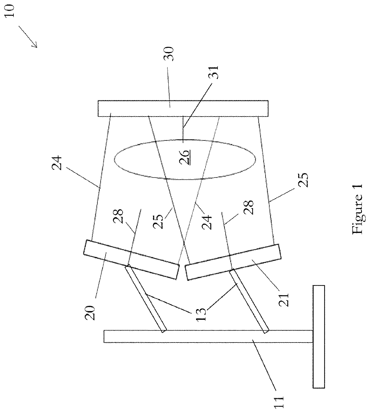
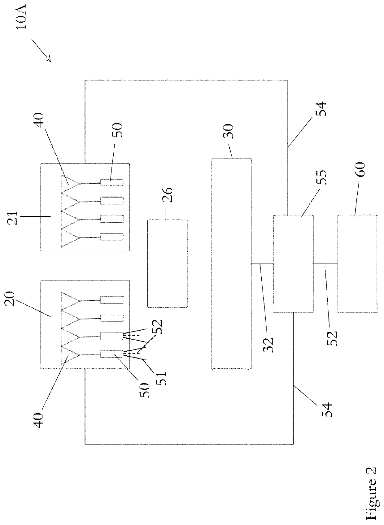
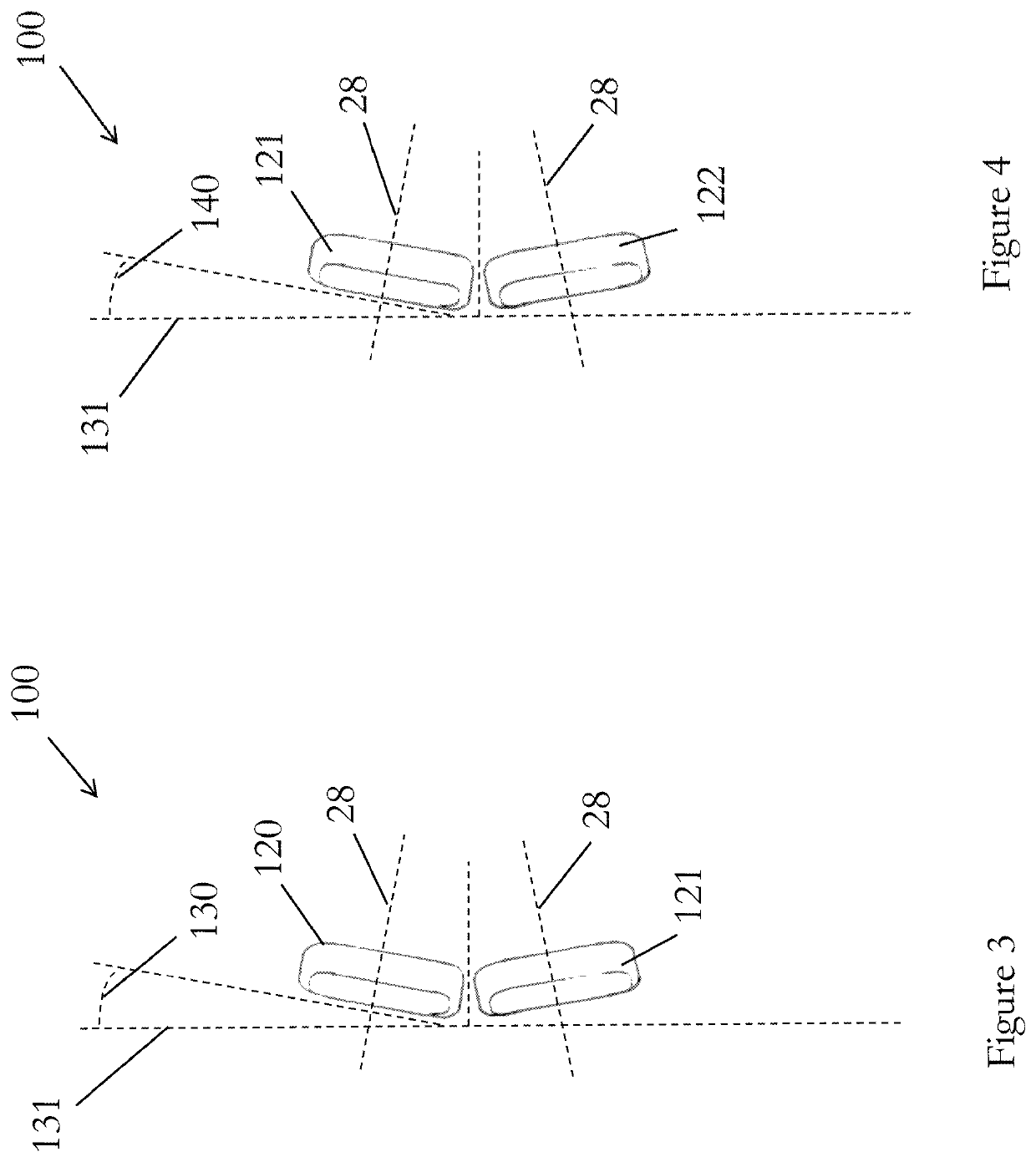
[0056]The present invention will be described with respect to certain drawings but the invention is not limited thereto but only by the claims. The drawings described are only schematic and are non-limiting. Each drawing may not include all of the features of the invention and therefore should not necessarily be considered to be an embodiment of the invention. In the drawings, the size of some of the elements may be exaggerated and not drawn to scale for illustrative purposes. The dimensions and the relative dimensions do not correspond to actual reductions to practice of the invention.

[0057]Furthermore, the terms first, second, third and the like in the description and in the claims, are used for distinguishing between similar elements and not necessarily for describing a sequence, either temporally, spatially, in ranking or in any other manner. It is to be understood that the terms so used are interchangeable under appropriate circumstances and that operation is capable in other s...


PUM


Abstract

An x-ray imaging device (10) comprising at least two substantially planar panels (20, 21), each panel comprising a plurality of x-ray emitters housed in a vacuum enclosure, wherein the at least two panels each have a central panel axis (28) and are arranged such that their central panel axes are non-parallel to one another, the device further comprising a panel retaining means and arranged such that the panel retaining means retains the at least two panels stationary in relation to an object during x-raying of the object.

Description

PRIORITY[0001]The present application is related to, and claims the priority benefit of, and is a 35 U.S.C. 371 national stage application of, International Patent Application Serial No. PCT / GB2018 / 053400, filed Nov. 23, 2018, which is related to, and claims the priority benefit of, Great Britain Patent Application Serial No. 1719599.1, filed Nov. 25, 2017. The contents of each of these applications are hereby incorporated by reference in their entirety into this disclosure.TECHNICAL FIELD[0002]The present invention relates generally to an x-ray imaging device and a method of producing an x-ray image and finds particular, although not exclusive, utility in medicine.BACKGROUND[0003]This invention is concerned with x-ray sources and more specifically with multiple x-ray sources. It is well known that x-ray tubes can be moved in a controlled fashion using gantries and computer controlled motorized stages so as to image an object from multiple angles and positions. It is also known that...

Claims


Application Information

Patent Timeline
IPC IPC(8): A61B6/00A61B6/02A61B6/06G21K1/02
CPCA61B6/547A61B6/025A61B6/06A61B6/4007A61B6/4085A61B6/4429G21K1/025A61B6/4476H01J2235/163H01J2235/068H01J2235/161G03B42/026H01J1/3048
Inventor MCKEAN, WESWELLS, STEVETRAVISH, GIL
Owner ADAPTIX
Why Eureka
Try Eureka