Method of tumor regression with VEGF inhibitors
- Summary
- Abstract
- Description
- Claims
- Application Information
AI Technical Summary
Problems solved by technology
Method used
Image
Examples
example 1
Regression of Established Tumors During VEGF-Trap Injection
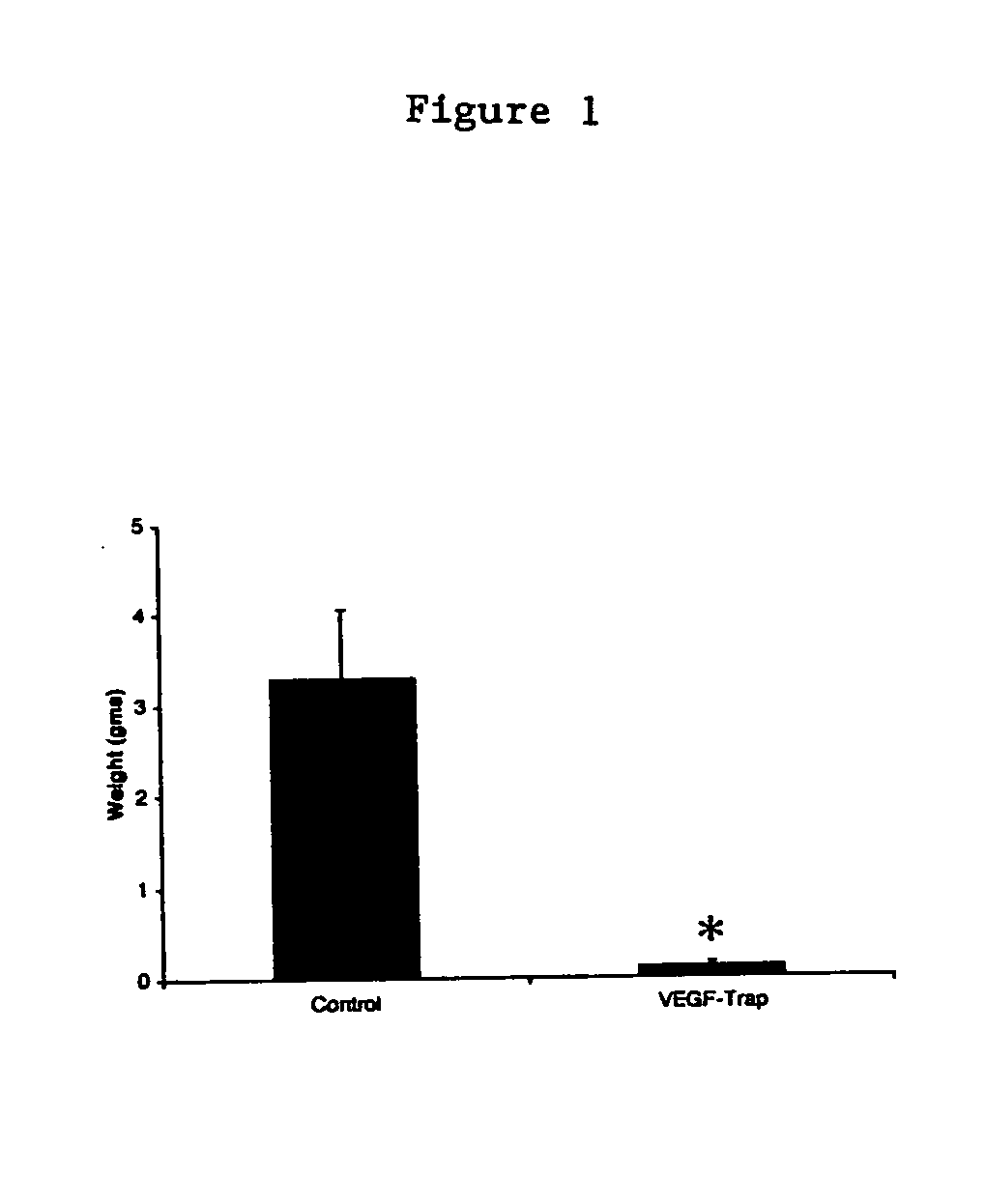
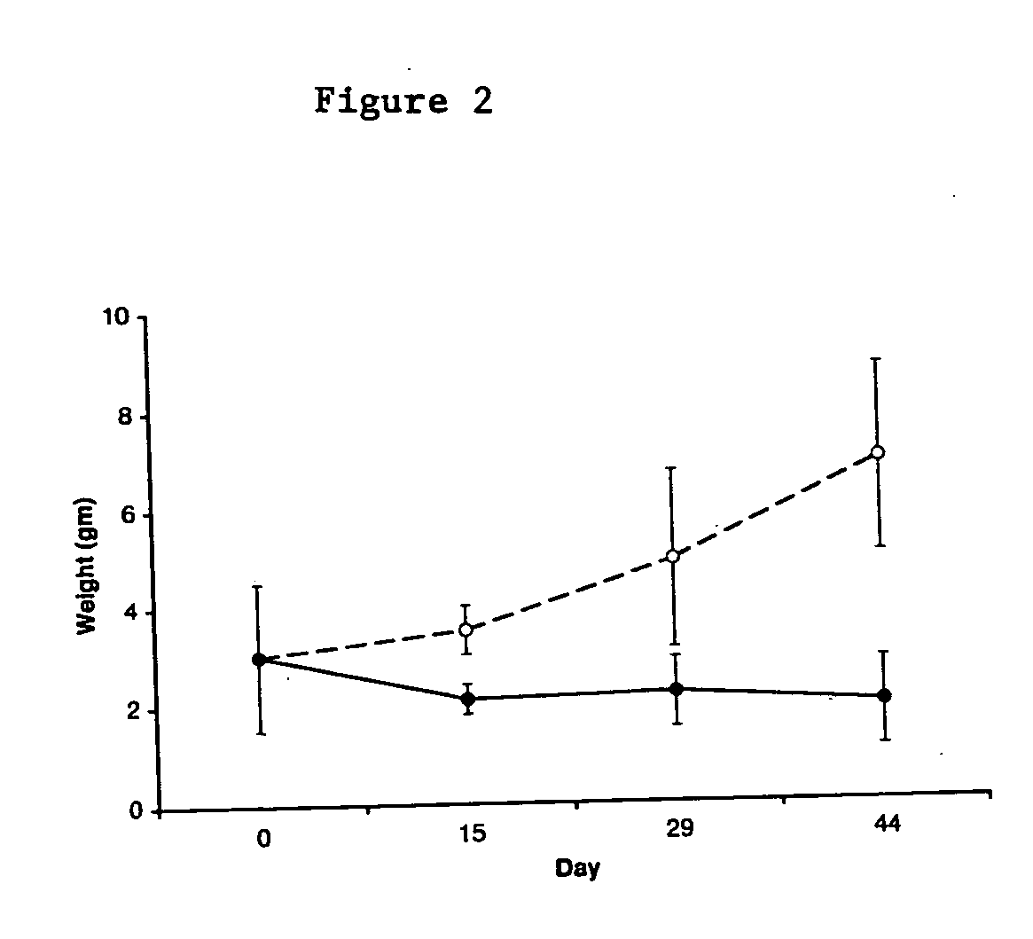
[0054] Xenograft Model. SK-NEP-1 Wilms tumor cells (American Type Culture Collection, Manassas, Va.), SY5Y neuroblastoma cells (American Type Culture Collection, Manassas, Va.) or HUH hepatoblastoma cells (HuH-6, RIKEN BioResource Center, Ibaraki, Japan) were maintained in culture with McCoy's 5A medium (Mediatech, Fisher Scientific, Springfield, N.J.), supplemented with 15% fetal bovine serum and 1% penicillin-streptomycin (Gibco, Grand Island, NY). Cells were grown at 37.degree. C. in 5% CO2 until confluent, harvested, counted with trypan blue staining, and washed and resuspended in sterile phosphate-buffered saline (PBS) at a concentration of 107 cells per milliliter. Xenografts were established in 4-6 week old female NCR nude mice (NCI-Frederick Cancer Research and Development Center, Frederick, Md.) by intrarenal injection of 106 cells from one of the following human cell lines: SK-NEP-1, SY5Y or hepatoblastoma cells an...
example 2
Involution of Existing Vasculature in Wilms Tumor During VEGF Trap Injection
[0059] Lectin perfusion. Prior to euthanasia, selected mice at each time point underwent intravascular injection of fluorescein-labeled Lycopersicon esculentum lectin (100 .mu.g in 100 .mu.l of saline, Vector Laboratories, Burlingame, Calif.) into the left ventricle. The vasculature was fixed by infusion of 1% paraformaldehyde (pH 7.4) in PBS, and then washed by perfusion of PBS, as described in Thurston et al. (1996) Am. J. Physiol. 271:H2547-2562.
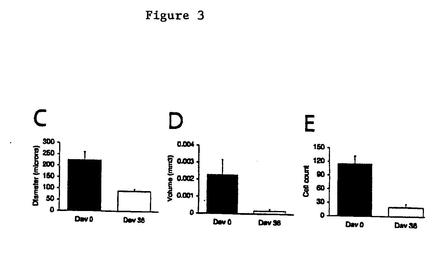
[0060] Digital Image Analysis. Digital images from the fluorescein-labeled lectin studies were acquired from a Nikon E600 fluorescence microscope (10.times. objective) with a Spot RT Slider digital camera (Diagnostic Instruments, Sterling Heights, Mich.) and stored as TIFF files. Quantitative assessment of angiogenesis and tumor vessel architecture was performed by computer-assisted digital image analysis as described by Wild et al. (2000) Microvasc. Res. 59:368-3...
example 3
Apoptosis in Endothelial and Vascular Mural Cells
[0068] PECAM-1, aSMA, and TUNEL double-staining. Apoptosis was determined by terminal deoxynucleotidyl transferase-mediated deoxyuridine triphosphate nick-end labeling (TUNEL) staining. Immunofluorescent double-staining for PECAM-1 / apoptosis and aSMA / apoptosis was performed on frozen sections using the ApopTag Red In Situ Apoptosis Detection Kit (Intergen Company, Purchase, N.Y.) and either rat anti-mouse PECAM-1 or anti-aSMA monoclonal antibody. A biotinylated secondary antibody was used in combination with fluorescein-labeled avidin to visualize endothelial and vascular mural cells, respectively. Slides were examined and photographed by fluorescence microscopy.
[0069] If VEGF-mediated signaling is critical to the survival of both the endothelial and vascular mural cells of mature tumor vessels, apoptosis should be detectable concurrently in both cell populations. Double labeling using the TUNEL assay combined with PECAM-1 and aSMA im...
PUM
| Property | Measurement | Unit |
|---|---|---|
| Size | aaaaa | aaaaa |
Abstract
Description
Claims
Application Information
 - R&D
- Intellectual Property
- Life Sciences
- Materials
- Tech Scout
- Unparalleled Data Quality
- Higher Quality Content
- 60% Fewer Hallucinations
Browse by: Latest US Patents, China's latest patents, Technical Efficacy Thesaurus, Application Domain, Technology Topic, Popular Technical Reports.
© 2025 PatSnap. All rights reserved.Legal|Privacy policy|Modern Slavery Act Transparency Statement|Sitemap|About US| Contact US: help@patsnap.com


