Method and apparatus for automatically detecting breast lesions and tumors in images

an automatic detection and breast technology, applied in the field of automatic detection of breast tumors and lesions in images, can solve the problems of fatigue and/or oversight of radiologist, inability to detect the presence of breast tumors and lesions, inaccurate manual interpretation, etc., to remove speckles, enhance contrast, and remove speckles.

Inactive Publication Date: 2005-02-03
RUTGERS THE STATE UNIV
View PDF13 Cites 94 Cited by
  • Summary
  • Abstract
  • Description
  • Claims
  • Application Information

AI Technical Summary

Benefits of technology

The present invention provides a method for automatically detecting breast tumors and lesions in images, including ultrasound, digital mammogram, and MRI images. The method comprises the steps of acquiring an image of a breast; filtering the image to remove speckle and enhance contrast; applying texture and intensity classifiers to each pixel of the image, the classifiers corresponding to probabilities of the pixel belonging to a lesion or tumor; determining a seed point in the image; growing a region of interest around the seed point; calculating directional gradients for each pixel in the image; determining boundary points of the region of interest using the directional gradients; and processing the boundary points with a deformable model to determine the presence or absence of a tumor or lesion in the image.
The present inv...

Problems solved by technology

However, manual interpretation is often inaccurate, and can fail to detect the presence of breast tumors and lesions.
Such misses have been attributed to the subtle nature of the visual findings, poor image quality, or fatigue and/or oversight by the radiologist.
However, such algorithms rely on manual delineation of the tumor boundaries, and do not automatically delineate such boundaries.
Further, automatically detecting tumors and extracting lesion boundaries in ultrasound images and digital mammograms is difficult due to the specular nature and the variance in shapes and appearances of sonographic lesions, as well as shadowing artifacts and tumor-like structures in the image, such as glandular tissue, coopers ligaments, and sub-cutaneous fat.
Such obstacles make it difficult to automatically determine the lesion area using con...

Method used


Image

  • Method and apparatus for automatically detecting breast lesions and tumors in images
  • Method and apparatus for automatically detecting breast lesions and tumors in images
  • Method and apparatus for automatically detecting breast lesions and tumors in images

Examples


Embodiment Construction

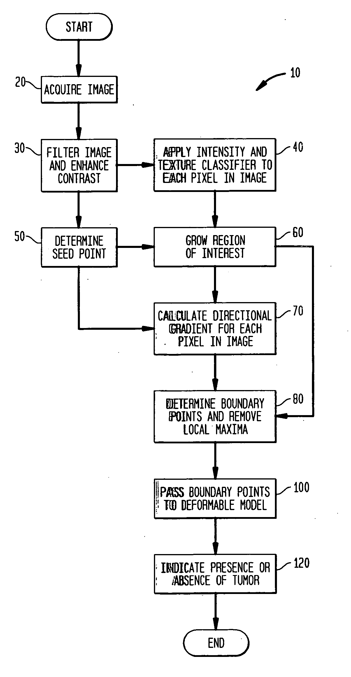
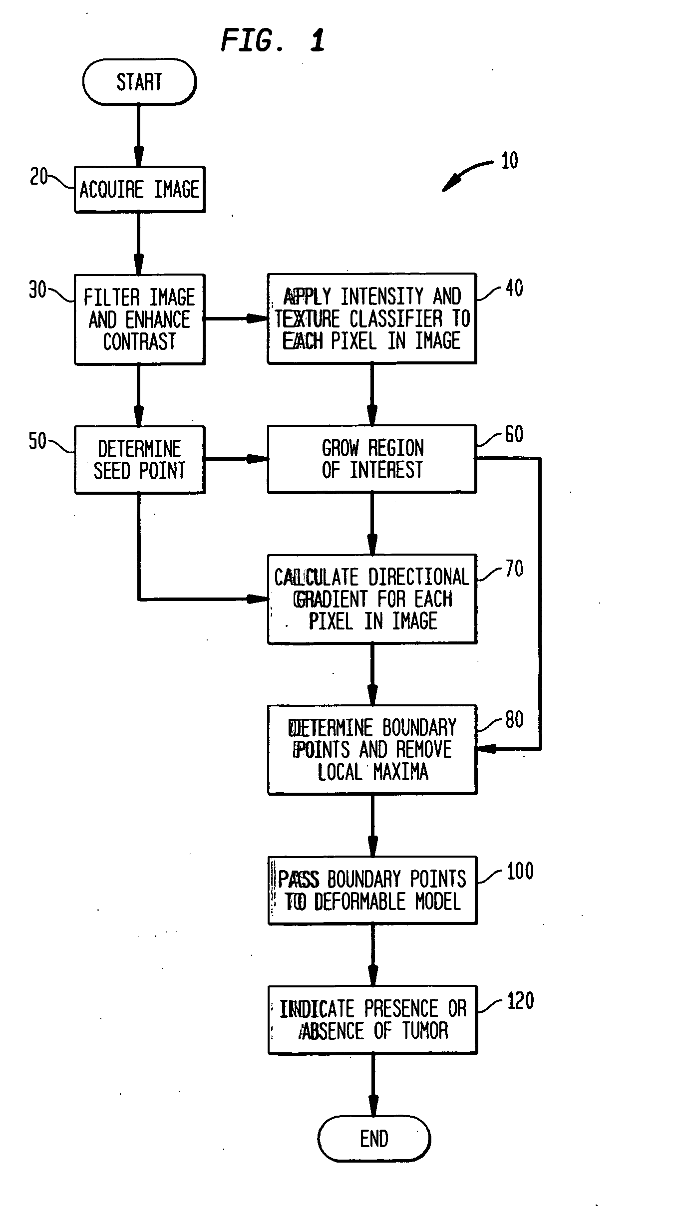
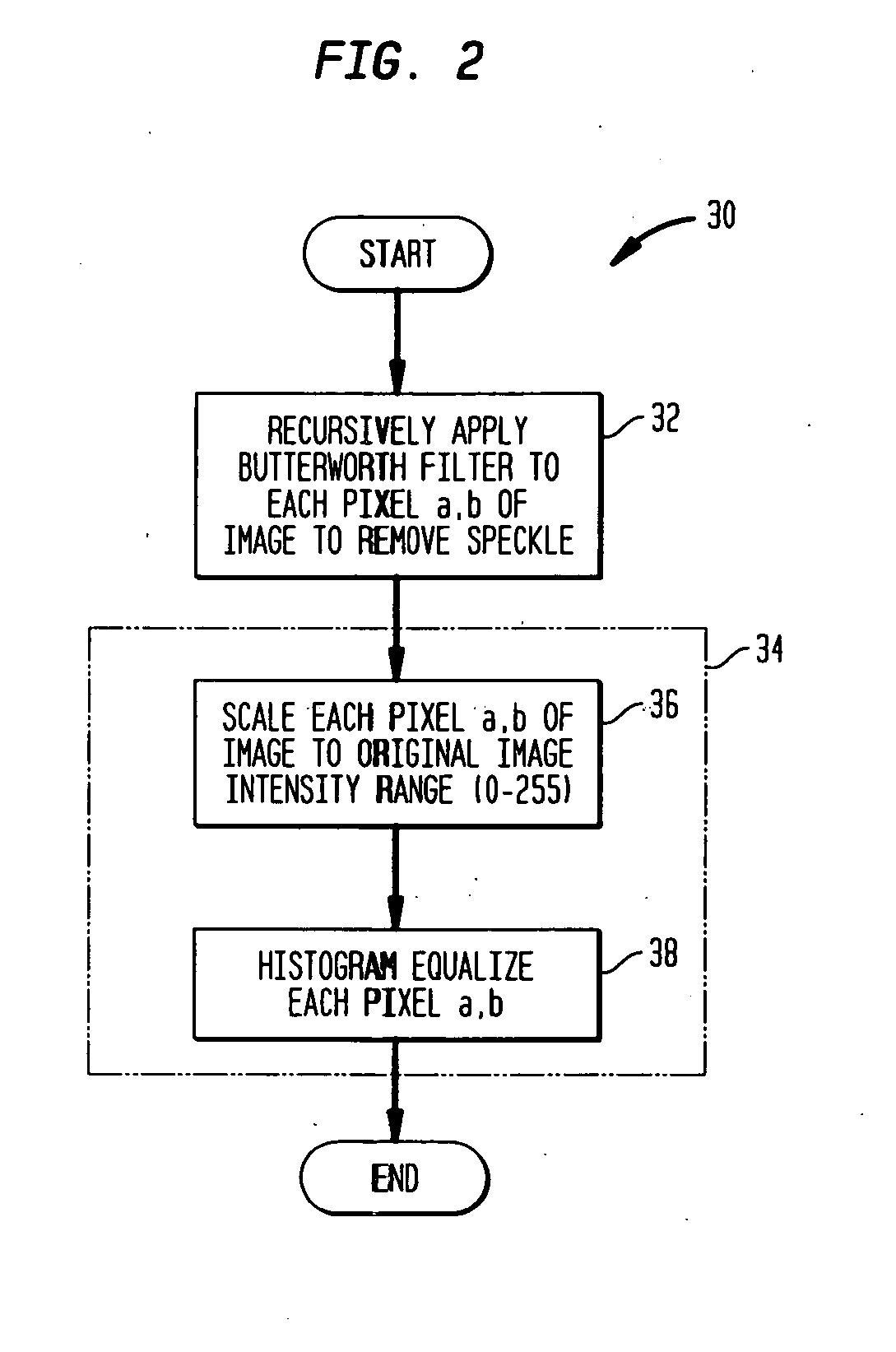
The present invention relates to a method and apparatus for automatically detecting breast tumors and lesions in images, including ultrasound, digital and analog mammograms, and MRI images. An image of a breast is acquired. The image is filtered and contrast of the image is enhanced. Intensity and texture classifiers are applied to each pixel in the image, the classifiers indicative of the probability of the pixel corresponding to a tumor. A seed point is identified within the image, and region of interest is grown around the seed point. Boundary points of the region of interest are identified. The boundary points are passed as inputs to a deformable model. The deformable model processes the boundary points to indicate the presence or absence of a tumor.

The present invention analyzes spatial distributions of various anatomic structures within a breast image, in addition to echogenicity of a lesion and its internal echo pattern, as three discriminating features for segmenting brea...


PUM


Abstract

A method and apparatus for automatically detecting breast tumors and lesions in images, including ultrasound, digital and analog mammograms, and MRI images, is provided. An image of a breast is acquired. The image is filtered and contrast of the image is enhanced. Intensity and texture classifiers are applied to each pixel in the image, the classifiers indicative of the probability of the pixel corresponding to a tumor. A seed point is identified within the image, and a region of interest is grown around the seed point. Directional gradients are calculated for each pixel of the image. Boundary points of the region of interest are identified. The boundary points are passed as inputs to a deformable model. The deformable model processes the boundary points to indicate the presence or absence of a tumor.

Description

BACKGROUND OF THE INVENTION 1. Field of the Invention The present invention relates to a method an apparatus for automatically detecting breast tumors and lesions in images. More specifically, the present invention relates to a method and apparatus for automatically segmenting, classifying, and detecting breast lesions in ultrasound, digital and analog mammogram, and magnetic resonance imaging (MRI) images. 2. Related Art Breast cancer is the most frequently diagnosed malignancy and the second leading cause of mortality in women. In the last decade, ultrasound imaging, along with digital mammography, has become the standard for breast cancer diagnosis. Mammography is the most effective method for early detection of breast cancer, and periodic screening of asymptomatic women reduces the mortality rate. Typically, the first step in breast cancer detection is a screening mammogram, which comprises a low-dose x-ray examination on asymptomatic women. This can be followed by a diagnos...

Claims


Application Information

Patent Timeline
IPC IPC(8): A61B5/055A61B8/08G06T5/00G06T7/00
CPCA61B5/055A61B5/7267A61B8/0825G06T7/0012G06T7/0081G06T7/0083G06T2207/30096G06T7/0089G06T2207/10088G06T2207/10132G06T2207/20156G06T2207/30068G06T7/0087G06T7/11G06T7/12G06T7/143G06T7/149G16H50/70
Inventor METAXAS, DIMITRIS N.MADABHUSHI, ANANT
Owner RUTGERS THE STATE UNIV
Why Eureka
Try Eureka