
Endoscope test device

Inactive Publication Date: 2005-03-03
LIGHTHOUSE IMAGING
View PDF7 Cites 23 Cited by
  • Summary
  • Abstract
  • Description
  • Claims
  • Application Information

AI Technical Summary

Benefits of technology

The invention provides an apparatus and methodology for quantitatively measuring the fitness of an endoscope for use in surgical procedures. The invention comprises a compact embedded processor which generates test patterns to a compact graphical display device, that are observed through an optical system that magnifies the image between the endoscope and the eye. The magnified images ease the burden on an operator's eye so that the measurements performed assess only the endoscope, not the combination of endoscope and eye. Specifically, the eye has a resolution of approximately 1 minute of arc. This is also approximately the resolution of the best quality endoscopes. The optical system magnifies the angular size of the image exiting the endoscope preferably by between 1.5-3×. Therefore, since the eye retains its 1 minute resolution, it needs to resolve only 1.5-3 minutes of arc at the least to assure that the endoscope, and not the observer, is measured.

Problems solved by technology

By their very nature, these devices are prone to failure from: Overstressing during surgery (particularly orthopedic) Carelessness during sterilization reprocessing Poor manufacturing design or workmanship Wear-and-tear after many sterilization cycles.
Improper repair, especially by unauthorized repair facilities
Unfortunately, there are currently no instruments available that provide hospitals with the clinically important and 0quantitative measurements to help determine whether an endoscope is fit for surgery.
None, however, meet the market's need for a cost-effective device that can be used in a clinical environment.
Its cost and complexity, however, make it most suitable to a manufacturing environment.
Unfortunately, the Endotester requires a custom computer system and appears to be complicated to use.
In addition, the Endotester is still in breadboard form and does not perform calibrated measurements, making the device of limited value in comparing test endoscopes to reference endoscopes.
The EQ-Test does not, however, provide the needed measurements for the assessment of image quality; it only assesses the photometric characteristics of endoscopes.
And, while interesting to the engineer and scientist, photometric characteristics of an endoscope are not particularly important clinically.
The limitation of performing endoscope quality tests without a computer frame-grab system is that when viewing an endoscope image with the eye, the limiting resolution of the endoscope is fairly well matched to the limiting resolution of the eye itself.
It is difficult, however, from a measurement standpoint.
For example, the eye at the end of a long day becomes tired and may not perform as well as in the morning.
Therefore, an endoscope measurement at the end of the day may appear worse than at the beginning since it was the eye that became defective and not the endoscope.

Method used


Image

  • Endoscope test device
  • Endoscope test device
  • Endoscope test device

Examples


Embodiment Construction

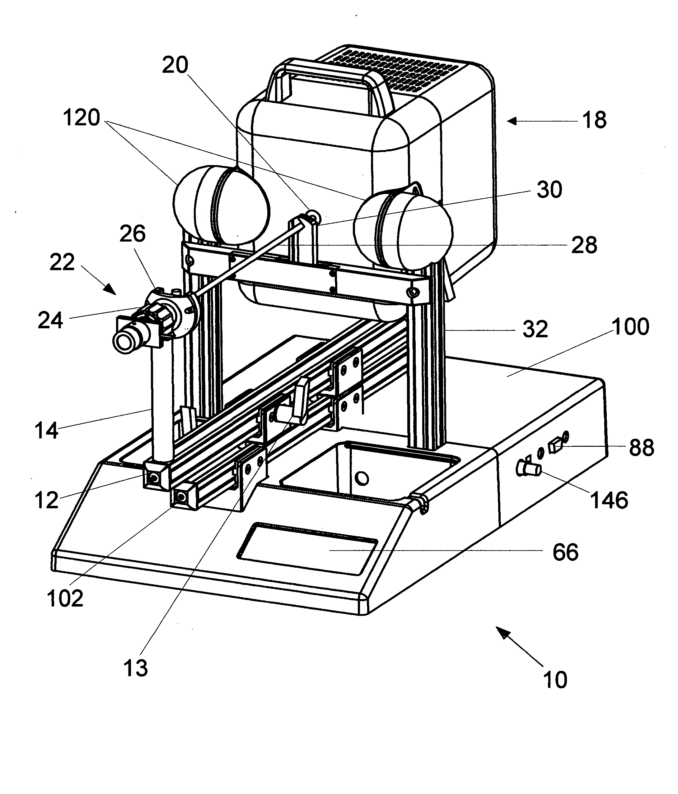
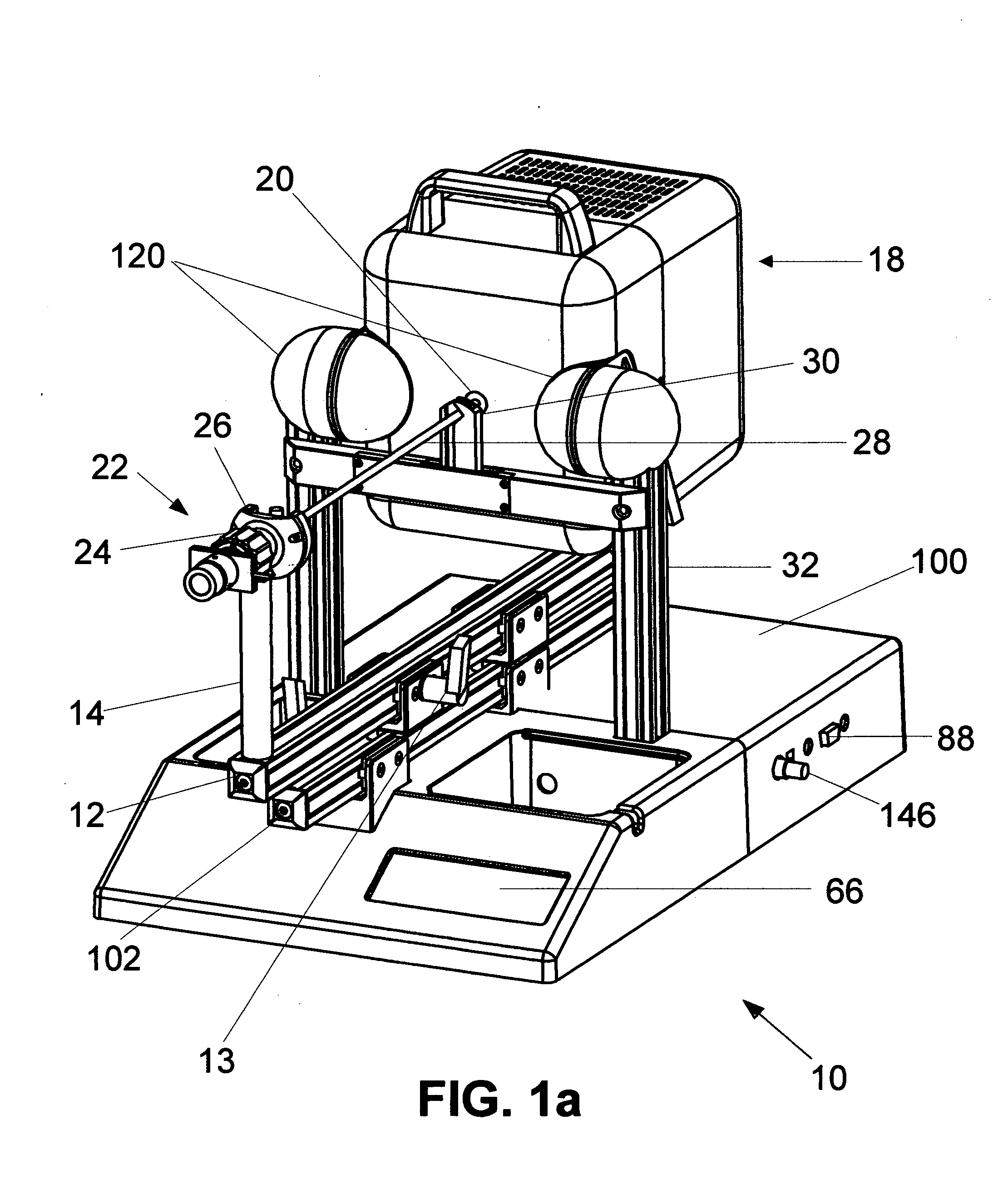
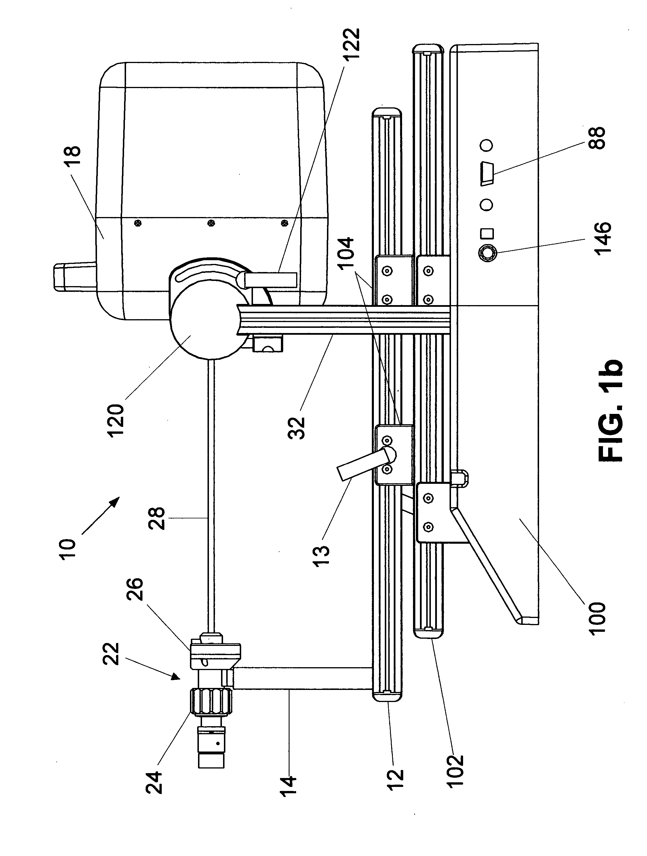
Reference is now made to FIGS. 1a-1c which show the inventive endoscope test system generally at 10. In a manner to be described, system 10 quantitatively measures the fitness of an endoscope for use in surgical procedures by generating user selectable predetermined test specific patterns on a compact graphical display device. The test patterns are observed through an optical system that magnifies the image between the endoscope and the eye, and the quality of the observed test patterns are scored by the observer and processed by system 10 that calculates a performance figure of merit indicating the endoscope's fitness for clinical use.

The major components of system 10 comprise a longitudinally extending rail 12 on which is mounted a slideable, vertically extending support 14. Above longitudinally extending rail 12 is a fixedly positioned cross frame member 16. Pivotally attached to cross frame member frame 16 is a housing 18 containing various electronics. Referring now to FIG. ...


PUM


Abstract

The invention uses optics and precision mechanisms to quantitatively assess the performance of medical endoscopes. By viewing standardized optical targets under well-controlled conditions, the optical quality of endoscopes can be easily determined in much the same fashion as getting one's eyes tested at the optometrist. The actual design, however, is much more complicated than your optometrist's eye chart and different-types of endoscopes from different manufacturers require customization of the target geometry, the viewing distance, and the viewing angle. Also, there are several optical characteristics beyond image sharpness that need to be assessed, requiring other unique test geometries, including contrast, distortion, and vignetting. Clinically significant measurements are performed with simplicity and cost-effectiveness through the use of relatively inexpensive optical components and an embedded processor and graphical display system.

Description

FIELD OF THE INVENTION This invention generally relates to apparatus and methodology for testing certain optical characteristics of medical endoscopes and borescopes and, more particularly, to apparatus and methodology for quantifying the quality of imaging through endoscopes and borescopes by providing tests for, including but not limited to, contrast, distortion, transmission, uniformity, and vignetting. The invention employs a CPU with a display device to generate specially formatted target images to an observer who then executes various scripted testing procedures that ultimately lead to a figure of merit for performance. BACKGROUND OF THE INVENTION The United States endoscopy market size in 1999 was $3.6 Billion. Visualization products, which include rigid endoscopes, light sources and video accessories, represent a 17%, or more, segment of this market valued at more than $600 Million. According to Stryker Corporation's 1999 Fact Book, the world endoscopy market approaches $6...

Claims


Application Information

Patent Timeline
IPC IPC(8): G01M11/00G01M11/02H04N5/225H04N17/00
CPCG01M11/00G01M11/0214H04N2005/2255H04N17/002G01M11/0264A61B1/00057H04N23/555
Inventor LEINER, DENNIS C.BUSH, MICHAEL
Owner LIGHTHOUSE IMAGING
Learn More