Apparatus and method for customized report viewer

a report viewer and applicator technology, applied in the field of medical image analysis and diagnosis, can solve the problem of time-consuming for radiologists to select different images

Inactive Publication Date: 2005-05-05
MERGE CAD
View PDF6 Cites 79 Cited by
  • Summary
  • Abstract
  • Description
  • Claims
  • Application Information

AI Technical Summary

Problems solved by technology

Given the large number of images, it is very time consuming for the radiologist to select different images for different reports.

Method used


Image

  • Apparatus and method for customized report viewer
  • Apparatus and method for customized report viewer
  • Apparatus and method for customized report viewer

Examples


Embodiment Construction

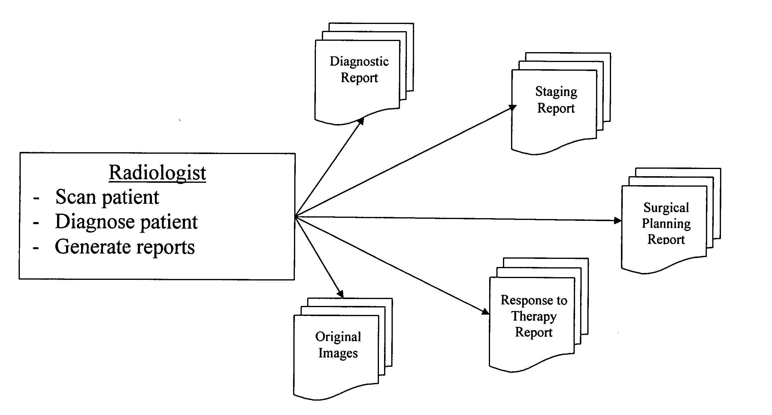
[0016] As will be discussed in further detail, the system described herein is directed to techniques for the automatic generation of custom reports from an initial set of image data. Various physicians have need for specific images and associated data. For example, the radiologist may evaluate certain images and the associate data to identify a volume of interest (VOI). Other physicians, such as a surgeon, may require a completely different set of images and associated data. Still other physicians, such as an oncologist, may have need for still another different set of images and associated data.

[0017] The types of procedures and reports needed for a patient varies depending on the state of the patient, and the physician interested in the results. There are reports needed for the initial diagnosis or screening of cancer. Once cancer has been identified, other reports are needed for staging, or determining the extent of disease. Based on the staging, patients may have surgery, in wh...


PUM


Abstract

A system for the automatic generation of custom report viewing utilizes imaging data and computer-aided detection technology to identify cancerous tumors. A typical collection of data from a patent's scan may include hundreds of images and associated data. The custom report viewer allows one physician, such as a radiologist, to analyze the data and prepare a report. The generated report may contain images, computed measurements, classifications based on a standard (such as the ACR BI-RADS for Breast MR), and locations relative to landmarks. Different physicians, such as an oncologist or a surgeon, may have need of differing images and supporting data for their own purposes. Each physician may select, in advance, custom selection criteria that are stored in association with that physician. A report generator module uses the stored selection criteria and report filtering to extract the images and supporting data specified by the particular physician. The system allows a surgeon to alter the selection criteria and obtain further images if necessary and permits the generation of multiple selection criteria by one physician for different purposes, such as surgical planning, therapy reporting, and the like.

Description

BACKGROUND OF THE INVENTION [0001] 1. Field of the Invention [0002] The present invention is directed generally to techniques for medical image analysis and diagnosis and, more particularly, to an apparatus and method for the generation of customized report viewing. [0003] 2. Description of the Related Art [0004] Breast cancer affects millions of individuals. In addition to breast self-examination, current medical advice includes periodic mammograms, which utilize conventional X-ray technology. Other forms of imaging, such as magnetic resonance imaging (MRI), are also known in the art. [0005] The initial data, which includes many images and may include additional laboratory test results, is evaluated by a physician. Typically, the radiologist evaluates images to determine whether a particular lesion is cancerous. The radiology must scan all of the patient reports, diagnose the patient and generate various reports. Given the large number of images, it is very time consuming for the r...

Claims


Application Information

Patent Timeline
Patent Type & Authority Applications(United States)
IPC IPC(8): A61B5/05G06F17/30G16H15/00G16H30/20
CPCG06F19/3487G06F19/321G16H15/00G16H30/20
Inventor DAW, SHAWNI L.BOISSERANC, JAMES J.
Owner MERGE CAD
Why Eureka
Try Eureka