
Segmented collimator assembly

Inactive Publication Date: 2005-05-19
GE MEDICAL SYST GLOBAL TECH CO LLC
View PDF5 Cites 7 Cited by
  • Summary
  • Abstract
  • Description
  • Claims
  • Application Information

AI Technical Summary

Problems solved by technology

Alignment of the scintillation detector array (often referenced as a pack) to the collimator in both x and z-directions (perpendicular plan to the direction of the x-ray beam) can be difficult.
Focal alignment of the plates can be difficult and costly.
Improper manufacturing can result in undesirable sensitivity to focal spot motion.
Manufacturing limitations, however, often present significant challenges to such larger collimator elements.
As collimator size increases, plate bow, dimensional accuracy, and alignment can all begin to negatively impact collimator and detector performance.
Although high precision manufacturing, fixturing, and precision features can at least partially address these concerns, they often are associated with undesirable cost increases.
The interface between adjoining modules, however, can become difficult to shield.
The edge between modules can become unshielded and negatively impact the resultant image produced by the imaging system.

Method used


Image

  • Segmented collimator assembly
  • Segmented collimator assembly
  • Segmented collimator assembly

Examples


Embodiment Construction

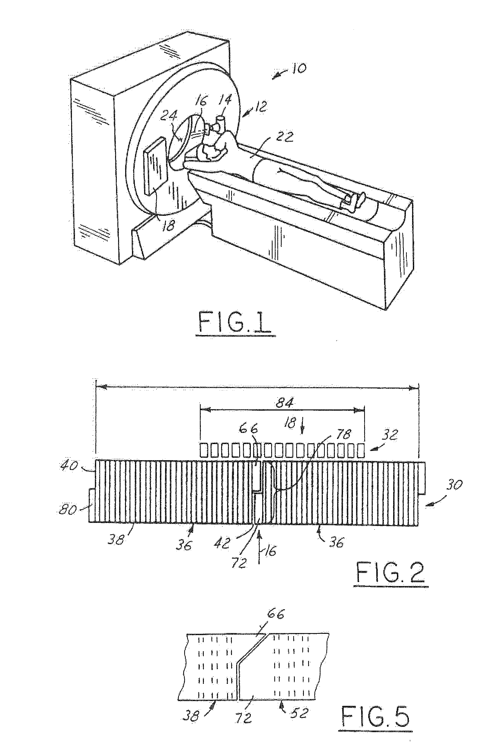
[0016] Referring now to FIG. 1, which is an illustration of a computed tomography (CT) imaging system 10 for use with the detector assembly 18 of the present invention. Although a particular CT imaging system 10 has been illustrated, it should be understood that the detector assembly 18 of the present invention can be utilized in a wide variety of imaging systems. The CT imaging system 10 includes a scanner assembly 12 illustrated as a gantry assembly. The scanner assembly 12 includes an x-ray source 14 for projecting a beam of x-rays 16 toward a detector assembly 18 positioned opposite the x-ray source 14. The detector assembly 18 senses the projected x-rays 16 that pass through an object, such as a medical patient 22. The detector assembly 18 produces an electrical signal that represents the intensity of an impinging x-ray beam and hence the attenuation of the beam 16 as it passes through the object of patient 22. Commonly, during a scan to acquire x-ray projection data, the scann...


PUM


Abstract

A detector assembly is provided comprising a collimator assembly. The collimator assembly comprises a first collimator segment having a first left end and a first right end. The first collimator segment includes a plurality of first segment longitudinal walls having a first segment depth. Each of the plurality of first segment longitudinal walls includes a first interlocking protrusion comprising only a portion of the first segment depth. The collimator assembly also includes a second collimator segment having a second left end and a second right end. The second collimator segment comprises a plurality of second segment longitudinal walls having a second segment depth. Each of the plurality of second segment longitudinal walls includes a second interlocking protrusion comprising only a portion of the second segment depth. Each of the second interlocking protrusions engages one of the first interlocking protrusions to form a continuous sidewall segment.

Description

BACKGROUND OF INVENTION [0001] The present invention relates generally to an x-ray beam collimator assembly, and, more particularly to a collimator assembly that can be manufactured as segments and assembled into larger collimators. [0002] Computed tomography has been utilized for a wide variety of imaging applications. One such category of applications is comprised of medical imaging. Although it is known that computed tomography may take on a wide variety of configurations within the medical industry, it commonly is based on the transmission of low energy rays (x-rays) through a body structure. These low energy rays are subsequently received and processed to formulate an image, often three-dimensional, of the body structure that can by analyzed by clinicians as a diagnostic aid. [0003] The reception of the low energy rays, such as x-ray photons produced in an x-ray tube, is often accomplished through the use of a device referred to as a scintillator camera or detector array. The s...

Claims


Application Information

Patent Timeline
IPC IPC(8): G01J1/00G01T1/164G01T1/24G21K1/02H01L25/00H01L27/00
CPCG21K1/025G01T1/1648
Inventor IKHLEF, ABDELAZIZ
Owner GE MEDICAL SYST GLOBAL TECH CO LLC
Features
  • Generate Ideas
  • Intellectual Property
  • Life Sciences
  • Materials
  • Tech Scout
  • Unparalleled Data Quality
  • Higher Quality Content
  • 60% Fewer Hallucinations
Learn More