
Apparatus and method for surgical planning and treatment monitoring

a technology of surgical planning and apparatus, applied in the field of surgical planning techniques, can solve problems such as positive margins

Inactive Publication Date: 2005-05-26
FUJIFILM HEALTHCARE CORP +1
View PDF2 Cites 197 Cited by
  • Summary
  • Abstract
  • Description
  • Claims
  • Application Information

AI Technical Summary

Benefits of technology

The present invention provides techniques for surgical planning and treatment monitoring using medical imaging techniques. The invention aims to improve the accuracy of surgical planning and reduce the need for additional surgeries to remove cancerous tissue. The invention uses a system that includes a central processing unit, memory, data storage, and imaging devices to process and classify imaging data. The system can detect and diagnose abnormalities automatically or assist the surgeon in identifying the location of the abnormality. The invention also provides a method for measuring the size of the abnormality and monitoring its growth or regression during treatment. Overall, the invention improves the accuracy of surgical planning and treatment monitoring, leading to better outcomes for patients.

Problems solved by technology

Unfortunately, this procedure often results in positive margins.

Method used


Image

  • Apparatus and method for surgical planning and treatment monitoring
  • Apparatus and method for surgical planning and treatment monitoring
  • Apparatus and method for surgical planning and treatment monitoring

Examples


Embodiment Construction

[0020] As will be discussed in further detail, the system described herein is directed to techniques for cataloging and measuring lesions or volumes of interest (VOI) for purposes of surgical planning and treatment monitoring. Although the techniques discussed herein use examples directed to evaluation of breast tumors, the techniques are more widely applicable to the evaluation of tissue for surgical planning purposes in general.

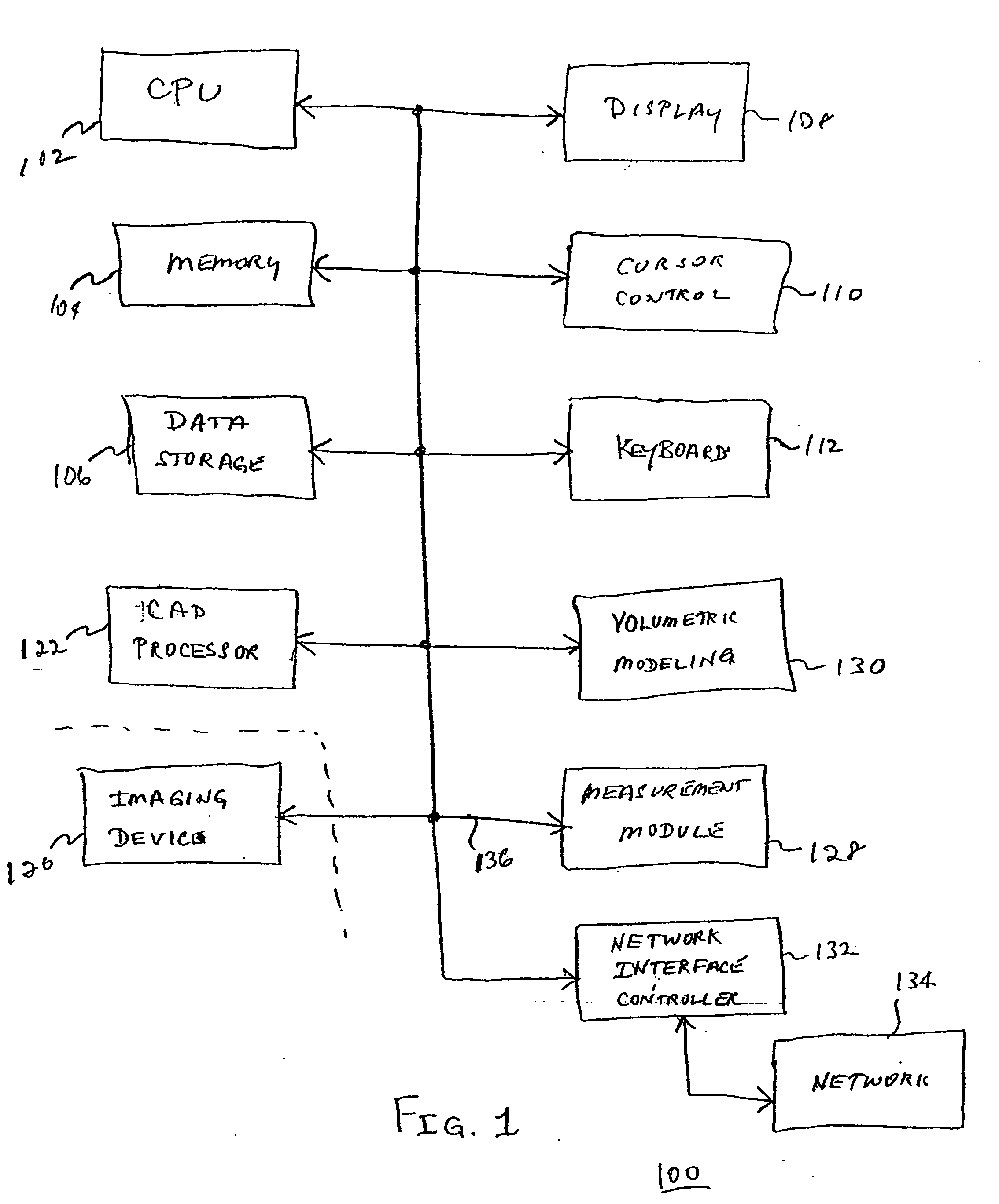
[0021]FIG. 1 is a functional block diagram of a system 100 constructed in accordance with the principles described herein. Many of the components of the system 100 are implemented as conventional computer components and need only be described briefly herein.

[0022] The system 100 includes a central processing unit (CPU 102) and a memory 104. The CPU 102 may be implemented as a microprocessor or part of a minicomputer or mainframe computer. The CPU 102 may be a conventional microprocessor chip, microcontroller, digital signal processor, or the like. Similar...


PUM


Abstract

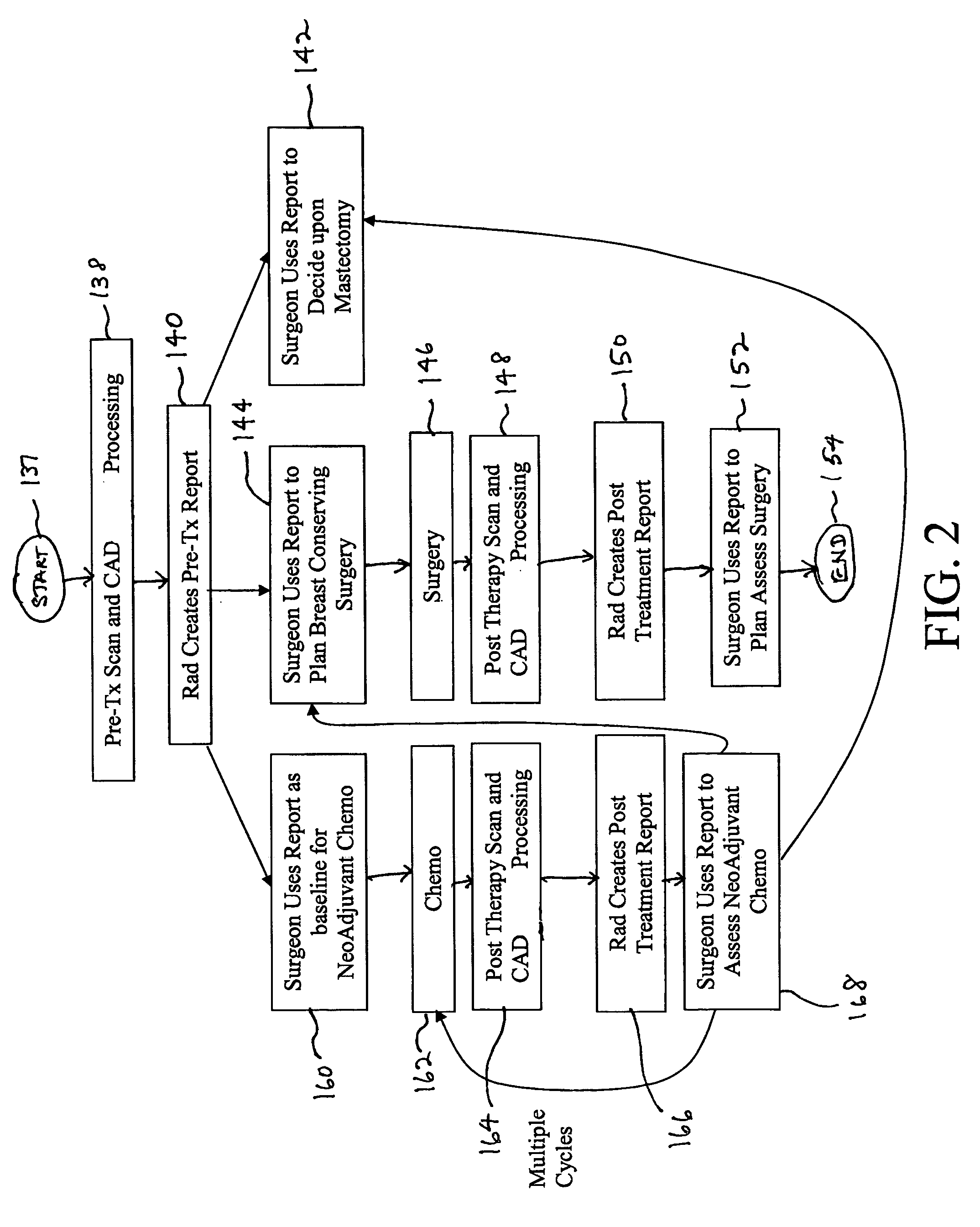
A system for surgical planning and therapeutic monitoring utilizes imaging data and computer-aided detection (CAD) technology to identify cancerous tumors. A pre-treatment report identifies all volumes of interest (VOIs) and provides data regarding the size and location of each VOI as well as volumetric data for use in surgical planning. The system can be used to monitor the progress of adjuvant chemotherapy or other non-surgical treatment and measures changes in tumor size and location. Post-treatment reports provide data regarding changes in tumor size and location as well as trend data to provide guidance to the physician.

Description

BACKGROUND OF THE INVENTION [0001] 1. Field of the Invention [0002] The present invention is directed generally to techniques for surgical planning and, more particularly, to an apparatus and method for surgical planning and treatment monitoring using medical imaging techniques. [0003] 2. Description of the Related Art [0004] Breast cancer affects millions of individuals. In addition to breast self-examination, current medical advice includes periodic mammograms, which utilize conventional X-ray technology. If lesions or tumors are discovered, the X-ray or mammogram is used to identify and locate the region. [0005] Conventional procedures for treatment include radiation and / or chemotherapy as well as surgical removal of the lesion. The surgical procedure may range from a lumpectomy to a mastectomy. Drug and radiation treatments are sometimes used pre-operatively to reduce or shrink the tumor size. [0006] In a typical lumpectomy, the surgeon uses X-ray to identify the region containi...

Claims


Application Information

Patent Timeline
Patent Type & Authority Applications(United States)
IPC IPC(8): A61B19/00G06T7/00
CPCA61B19/50A61B19/52G06F19/3437G06T2207/30068G06Q50/24G06T7/0012G06F19/3487A61B90/36A61B34/10G16H50/50G16H15/00G16H20/40
Inventor WOOD, CHRIS H.BOISSERANC, JAMES J.
Owner FUJIFILM HEALTHCARE CORP
Learn More