Automated centerline detection algorithm for colon-like 3d surfaces

a technology of colonlike 3d surface and automatic detection algorithm, which is applied in the field of automatic centerline detection algorithm for colonlike 3d surface, can solve the problems of challenging further processing of images such as detecting the centerline of colon, and achieve the effect of accurate ring profile of colonlike surfa

Active Publication Date: 2005-06-02
US DEPT OF HEALTH & HUMAN SERVICES
View PDF6 Cites 25 Cited by
  • Summary
  • Abstract
  • Description
  • Claims
  • Application Information

AI Technical Summary

Benefits of technology

[0018] A virtual non-invasive colonoscopy of a colon like surface includes processing computer tomographic image slices of the patient. The procurement of a three dimensional image of the colon like surface of vertex points, each vertex point having a discrete point identifier and three dimensional position information is presumed. The three dimensional position information of all vertex's neighbors is averaged in a shrinking procedure to contract the three dimensional image proximate to a major axis of the colon-like surface, with the overall length of the colon along the major axis remaining substantially unchanged. Evenly spaced points are taken th

Problems solved by technology

Assuming the presence of such an image, further processing of the image su

Method used


Image

  • Automated centerline detection algorithm for colon-like 3d surfaces
  • Automated centerline detection algorithm for colon-like 3d surfaces
  • Automated centerline detection algorithm for colon-like 3d surfaces

Examples


Embodiment Construction

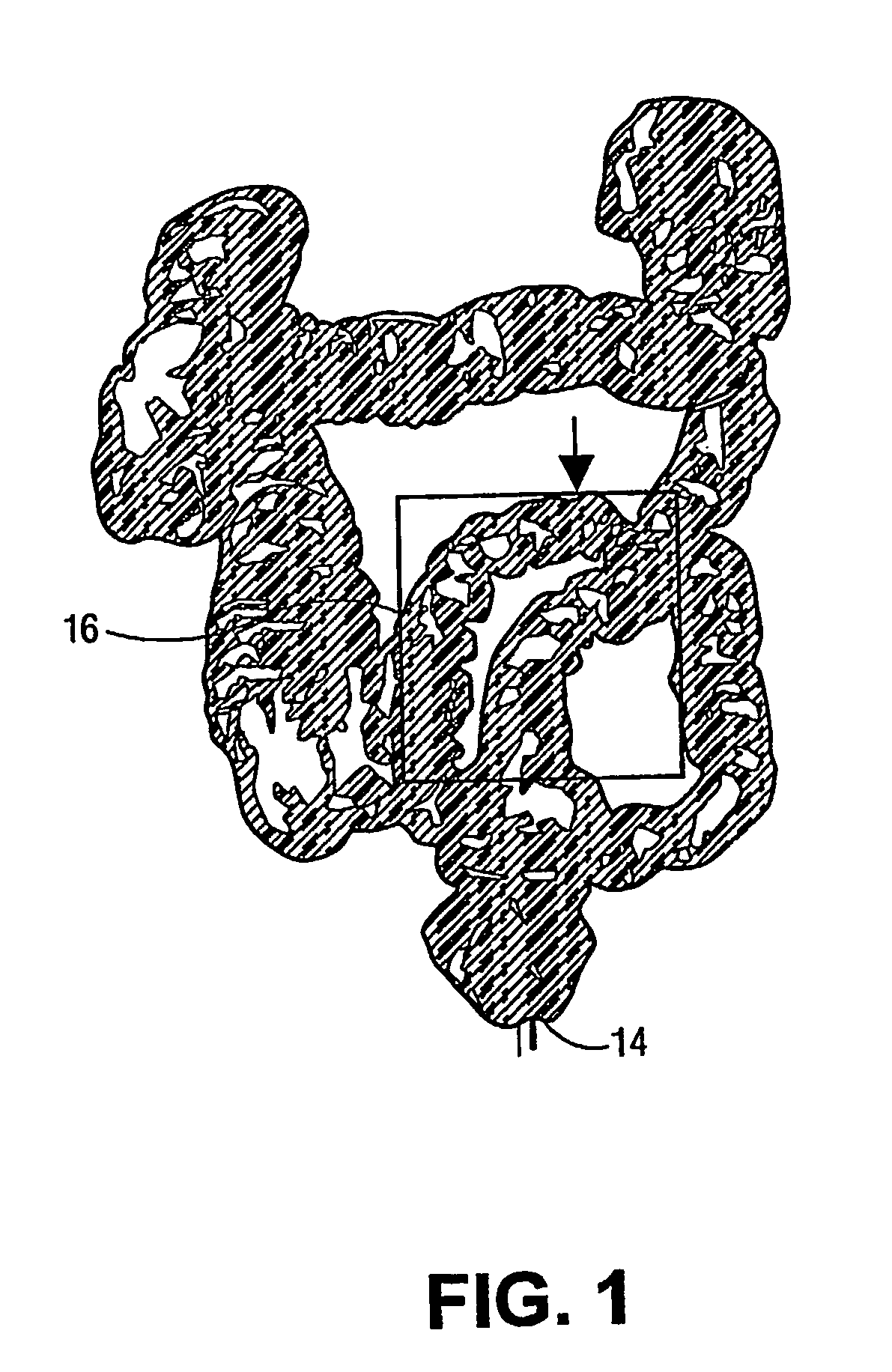
[0048] Referring to FIG. 1, we assume the acquisition of a 3-D image of the colon like surface such as that disclosed in Summers et al. U.S. Pat. No. 6,246,748 issued Jun. 12, 2001 and entitled Method for Segmenting Medical Images and Detecting Surface Anomalies in Anatomical Structures. We are using the results of the module which process the CT slices and computes the 3D surface of the colon in accordance with that disclosure. In this view, the anal verge 14 and the cecum 16 can be seen at either end of the colon.

[0049] Having acquired such an image, our problem is: given the 3D surface of the colon we want to find an (ordered) set of 3D points which define the colon 's centerline.

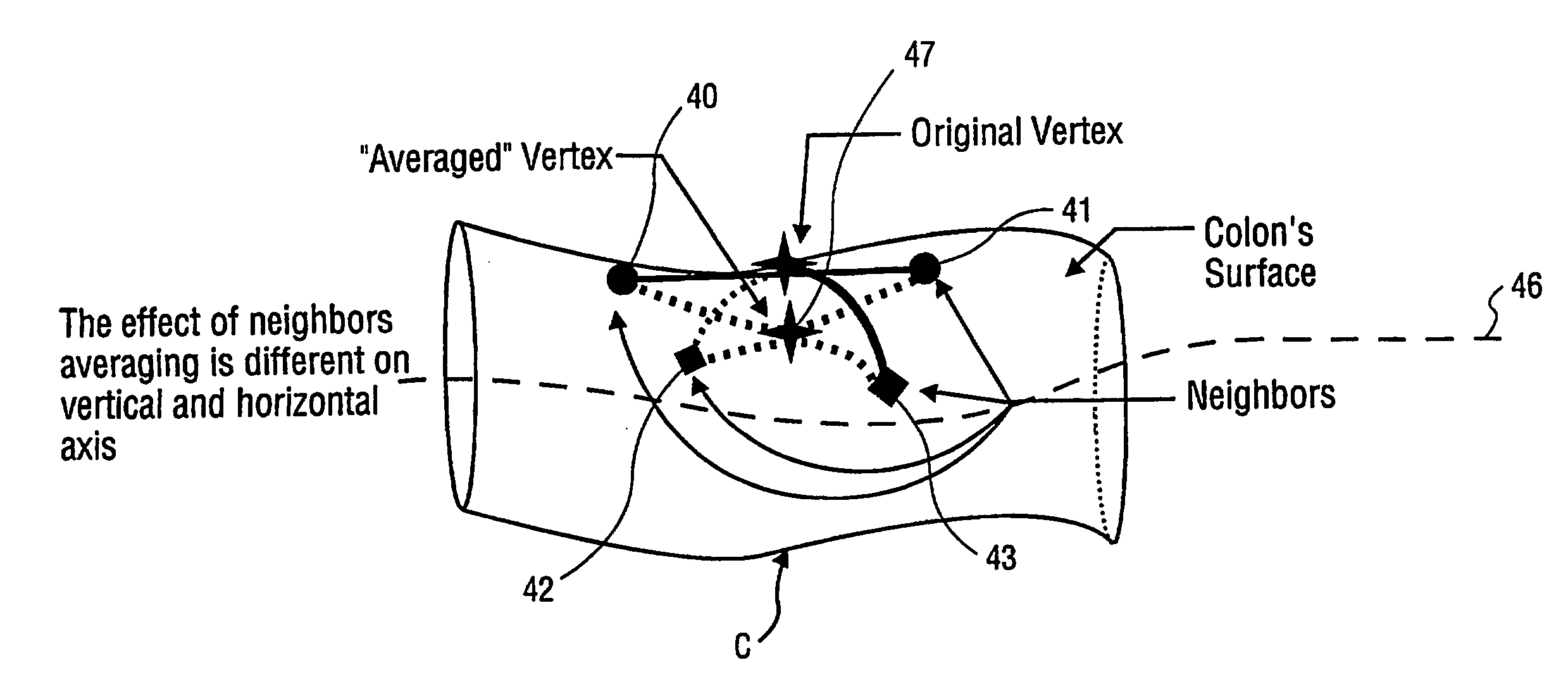
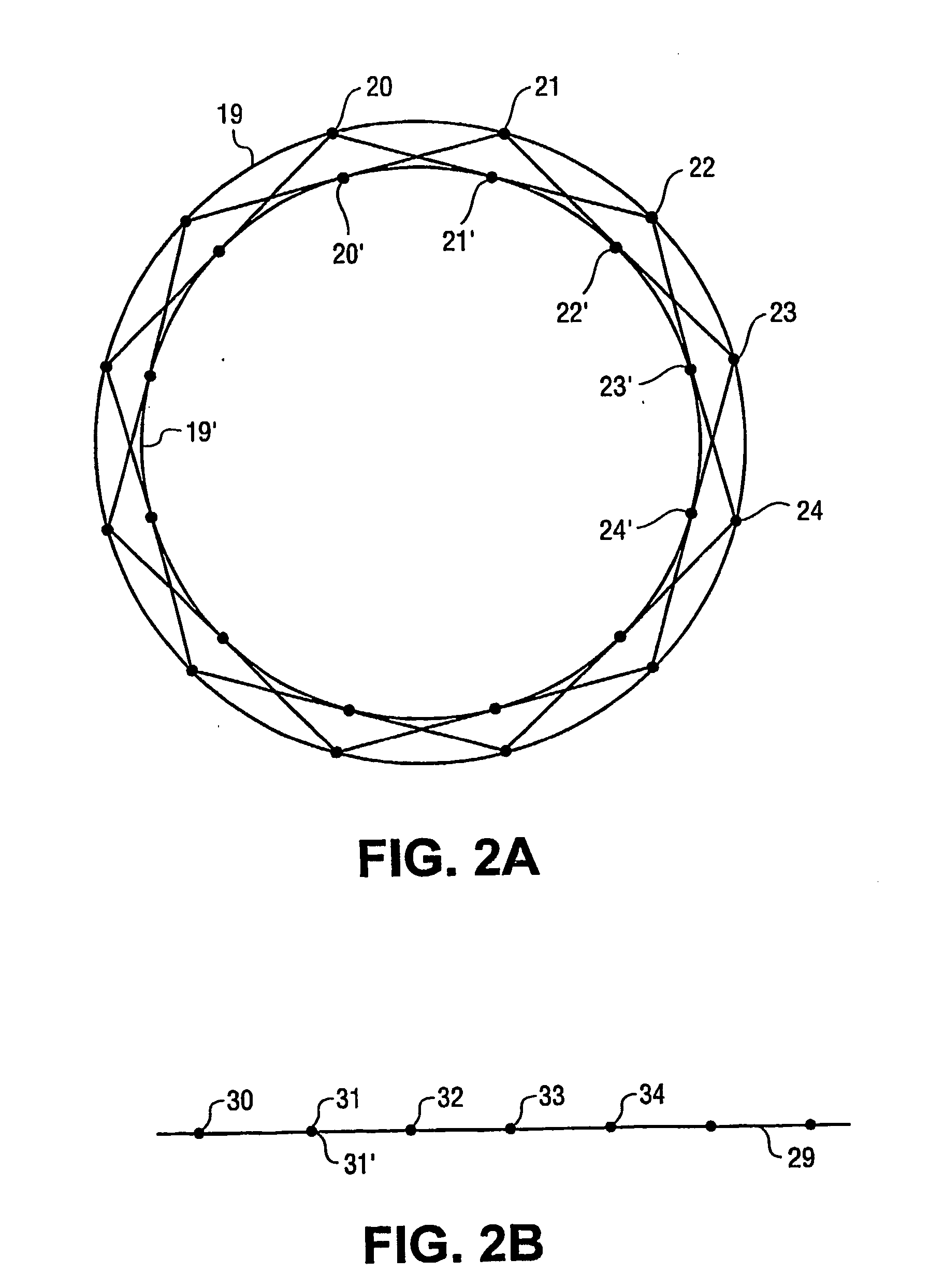
[0050] The method involves basic steps: [0051] 1) Compute a shrunken version of the colon. Basically the distances between vertices are iteratively averaged, the result being a shrunken colon. The shrunken colon is very thin and almost as long as the original colon. [0052] 2) Model the shrunken colon b...


PUM


Abstract

A three dimensional image of the colon like surface is processed to determine at least its ring structure. The image is composed of vertex points, each vertex point having a discrete point identifier and three dimensional position information. The three dimensional position information is averaged in a shrinking procedure to contract the three dimensional image proximate to a major axis of the colon-like surface. Evenly spaced points are taken through the shrunken colon like surface and connected to form a curve. Planes are generated at intervals normal to the curve to split the shrunken colon like surface into image segments. By mapping these image segments back to the original image through their discrete point identifiers, an accurate ring profile of the colon like surface can be generated.

Description

[0001] This invention relates to virtual colonoscopy and includes processing computer tomographic image slices of the patient. The disclosure assumes the procurement of a three dimensional image of the colon like surface of vertex points distributed along an elongate major axis, each vertex point having a discrete point identifier and three dimensional position information. In this disclosure, an accurate ring profile of the colon like surface can be generated. Utilizing the accurate ring profile, reconstruct the 3D surface of the colon, serially view the colon cross-section for anomalies such as polyps and thus provide both non-invasive colon screening together with the positional information needed for follow up conventional colonoscopy. BACKGROUND OF THE INVENTION [0002] Virtual Colonoscopy is a relatively new field in medicine and consists in processing the computer tomographic (CT) image planes or “slices” of the patient rather than the traditional and invasive colonoscopic ins...

Claims


Application Information

Patent Timeline
IPC IPC(8): G06K9/00G06T5/00G06T19/00
CPCG06T7/0083G06T7/0091G06T7/60G06T19/00G06T2210/41G06T2207/10081G06T2207/20044G06T2207/30028G06T2207/30172G06T2200/04G06T7/12G06T7/155
Inventor IORDANESCU, GHORGHESUMMERS, RONALD M.CEBRAL, JUAN RAUL
Owner US DEPT OF HEALTH & HUMAN SERVICES
Why Eureka
Try Eureka