Biopsy localization method and device

a biopsy and localization technology, applied in the field of biopsy localization methods and devices, can solve the problems that surgeons often have difficulty in determining the precise relationship of previously excised tissue to the surgical wound

Inactive Publication Date: 2005-02-10
DEVICOR MEDICAL PROD
View PDF90 Cites 104 Cited by
  • Summary
  • Abstract
  • Description
  • Claims
  • Application Information

AI Technical Summary

Benefits of technology

[0006] The present invention is directed to a biopsy localization method and device which uses a locatable bioabsorbable element left at the biopsy site so that if testing of the biopsy sample indicates a need to do so, the biopsy site can be relocated by finding the bioabsorbable element. Th

Problems solved by technology

For example, during a wide surgical excision for cancer diagnosed by a recent surgical excisional biopsy, surgeons fre

Method used


Image

  • Biopsy localization method and device
  • Biopsy localization method and device
  • Biopsy localization method and device

Examples


Embodiment Construction

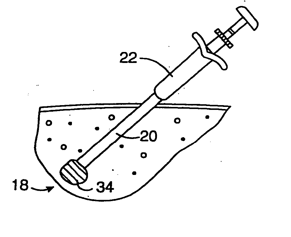
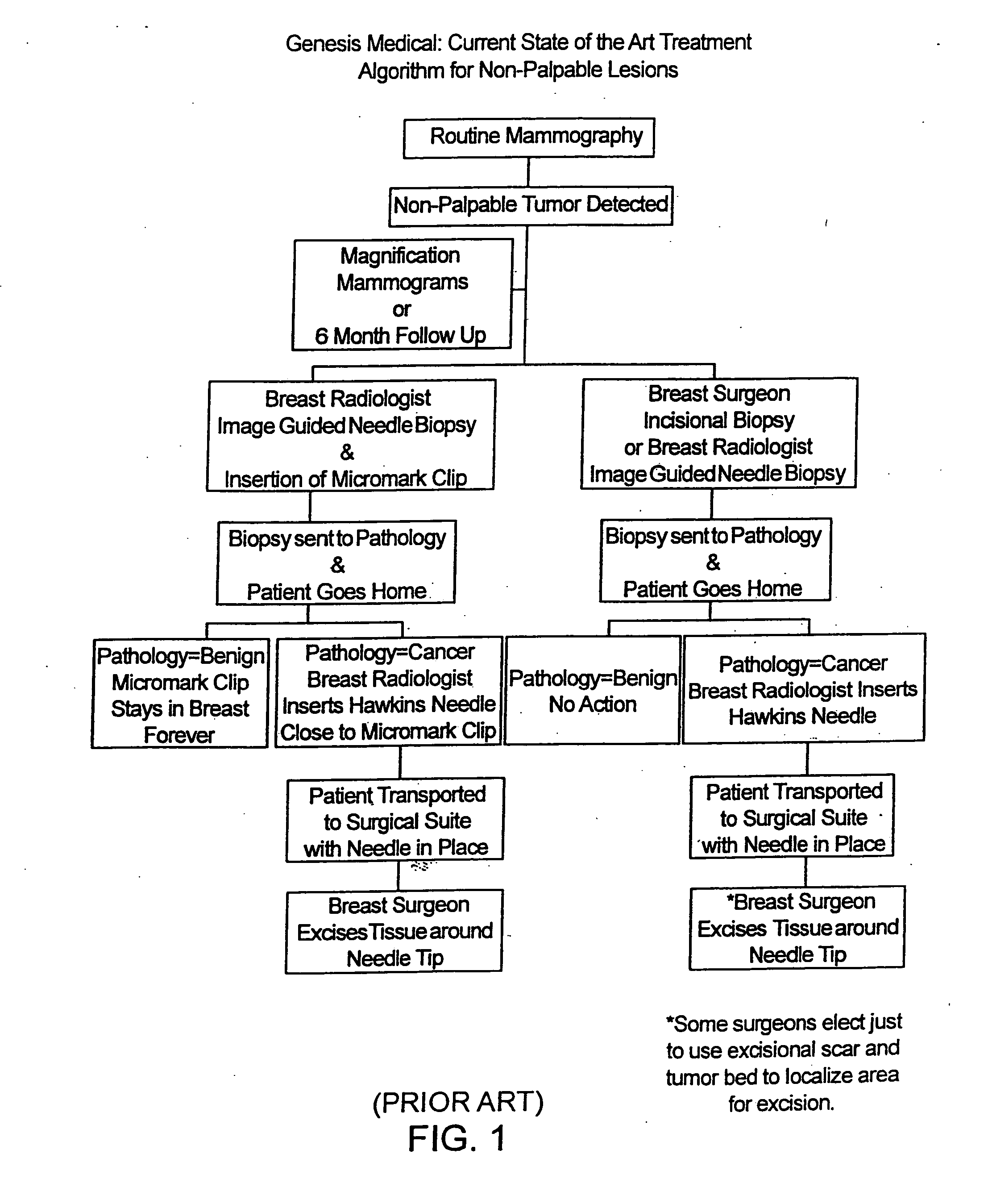
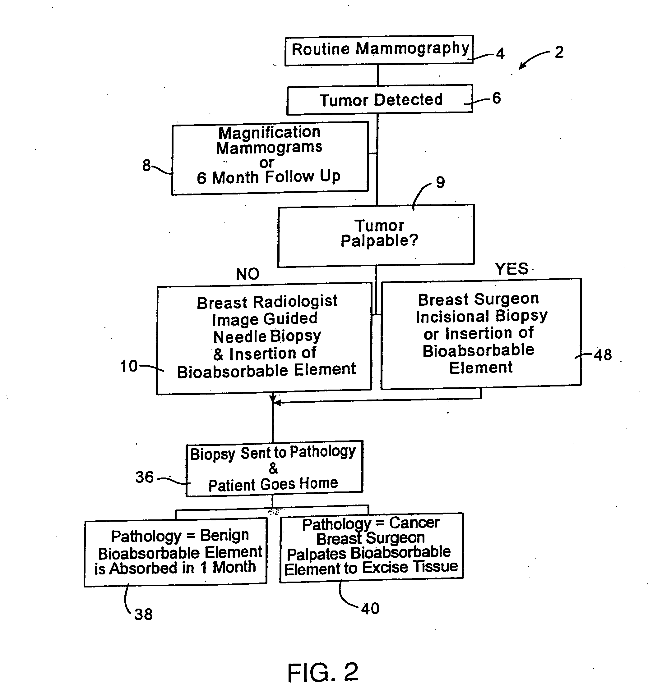
[0023]FIG. 2 illustrates a treatment algorithm 2 according to the present invention. As a result of a routine mammography 4, a tumor or other abnormality may be detected as at 6. The typical response will often include additional magnification mammograms or a follow-up mammogram scheduled for some time in the future, such as six months. This is indicated at 8. If the tumor is not palpable, see 9, an image guided needle biopsy by a breast radiologist is typically conducted as at 10. Image guided needle biopsies can be done in a number of ways. Presently, stereotactic (x-ray) and ultrasound guided needle biopsies are commonly used, primarily because of their accuracy, speed and minimal trauma to the patient. Stereotactic needle biopsies typically use a stereotactic table, such as made by Fisher or Lorad, which provides mammography (x-ray) guidance to a biopsy needle assembly. Ultrasound guided biopsies can be conducted with any one of a number of commercially available instruments. An...


PUM


Abstract

Methods for localizing a biopsy site are disclosed. The method includes taking a tissue sample from a biopsy site and positioning a detectable, bioabsorbable element at the biopsy site at the time that the tissue sample was taken. The tissue sample is then tested. The biopsy site is then relocated by finding the bioabsorbable element. The bioabsorbable element may be made of collagen, gelatin, cellulose, polylactic acid, and/or polyglycolic acid. The detectable bioabsorbable element may be relocated using ultrasound or mammography. The bioabsorbable element may also swell upon contact with body fluid.

Description

[0001] This application is a continuation of U.S. application Serial No. 10 / 839,112, filed May 4, 2004, which is a continuation of U.S. application Ser. No. 10 / 027,157, filed Dec. 20, 2001, now issued as U.S. Pat. No. 6,730,042, which is a continuation of U.S. application Ser. No. 09 / 900,801, filed Jul. 6, 2001, now issued as U.S. Pat. No. 6,699,205, which is a continuation of U.S. application Ser. No. 09 / 336,360, filed Jun. 18, 1999, now issued as U.S. Pat. No. 6,270,464, which application claims the benefit of the following Provisional patent applications: Biopsy Localization Device, Application No. 60 / 090,243, filed Jun. 22, 1998; Biopsy Localization and Hemostasis Device, Application No. 60 / 092,734, filed Jul. 14, 1998; Device and Method of Biopsy Localization and Hemostasis, Application No. 60 / 114,863, filed Jan. 6, 1999; and Device and Method of Biopsy Localization, Hemostasis & Cancer Therapy, Application No. 60 / 117,421, filed Jan. 27, 1999, all of the which are herein expres...

Claims


Application Information

Patent Timeline
IPC IPC(8): A61B6/00A61B10/02A61B10/00A61B17/34A61B19/00
CPCA61B6/502A61B10/02A61B17/3421A61B17/3468A61B2019/5487A61B2017/00004A61B2019/5408A61B2019/5462A61B19/54A61B2090/3962A61B2090/3908A61B2090/3987A61B90/39
Inventor FULTON, RICHARD E.DUBRUL, WILLIAM RICHARD
Owner DEVICOR MEDICAL PROD
Why Eureka
Try Eureka