Anti-CD40 antibodies capable of blocking B-cell activation

a technology of anti-cd40 antibodies and b-cell activation, applied in the field of new anti-cd40 antibodies capable of blocking b-cell activation, can solve the problem of inability to inhibit the b-cell response, and achieve the effect of preventing the growth or differentiation of b cells

Inactive Publication Date: 2005-08-04
NOVARTIS VACCINES & DIAGNOSTICS INC
View PDF18 Cites 1 Cited by
  • Summary
  • Abstract
  • Description
  • Claims
  • Application Information

AI Technical Summary

Problems solved by technology

However, the anti-CD40 antibodies known in the art stimulate B cells but are incapable of inhibiting the B-cell response.

Method used


Image

  • Anti-CD40 antibodies capable of blocking B-cell activation
  • Anti-CD40 antibodies capable of blocking B-cell activation
  • Anti-CD40 antibodies capable of blocking B-cell activation

Examples


example 1

Making Monoclonal Antibodies to B7 and CD40

A. PCR Cloning of CD40 and B7

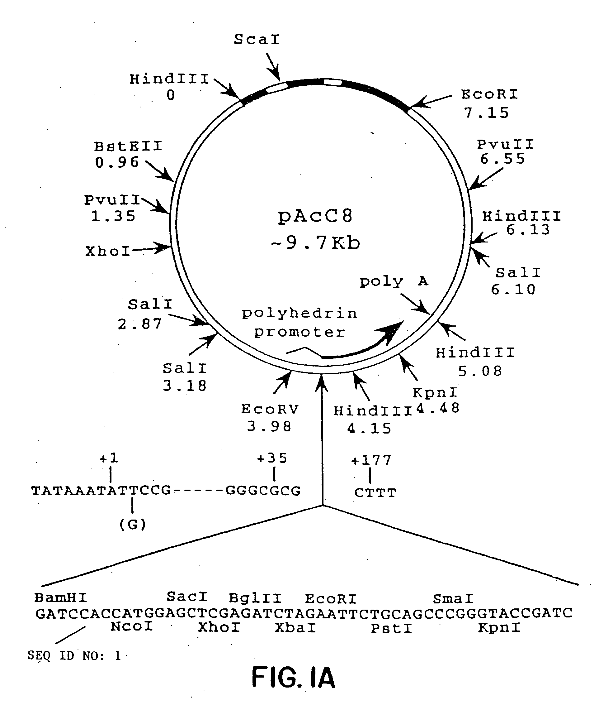
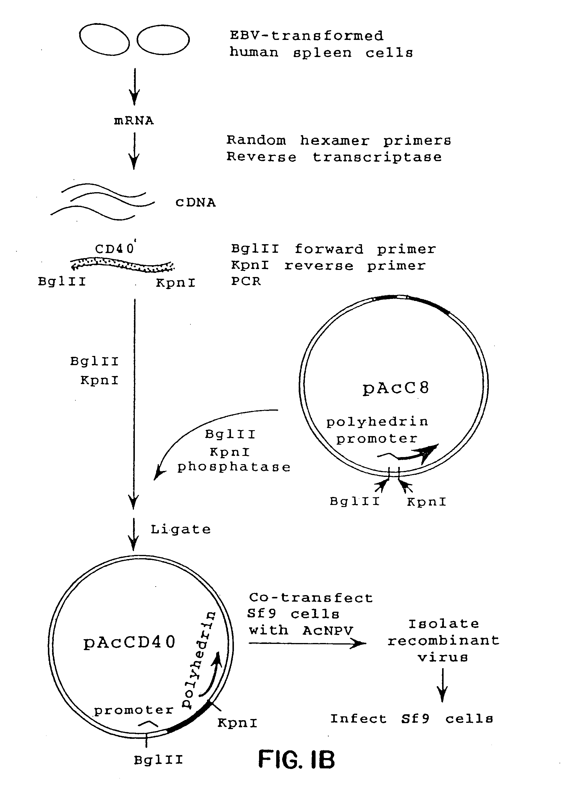
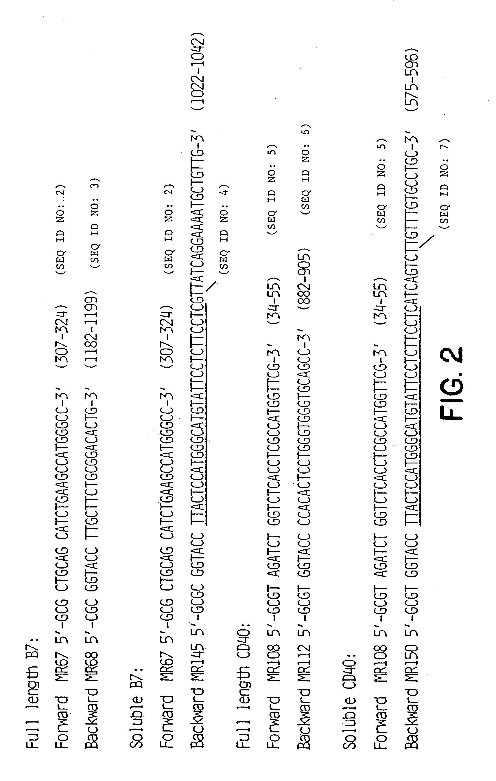
[0063] RNA was isolated from a population of EBV-transformed human spleen cells essentially as described by Chirgwin et al., Biochemistry (1979) 17:5294. In brief, the cells were washed twice with phosphate buffered saline (PBS) and lysed in 5 M guanidinium thiocyanate in the presence of 0.7 M 2-mercaptoethanol. The cell lysate was layered on a discontinuous CsCl gradient (Chirgwin et al.) and centrifuged for 16 hours at 26,000 rpm in a Beckman SW28 rotor. The RNA was recovered by dissolving the pellet in DEPC-treated H2O. The RNA was precipitated with ethanol once, resuspended in DEPC-treated H2O, and stored at −70° C.

[0064] Total RNA (10 μg / reaction) was converted to cDNA using random hexamer priming in 50 μl reaction buffer containing 500 units MLV-RT (Bethesda Research Laboratories, Bethesda, Md.), 5 μM random hexamer (Pharmacia, Piscataway, N.J.), 1 mM DTT, dNTP mix (0.5 mM each), 10 mM Tris-HCL pH 8.3,...

example 2

Costimulation of B-Cell Proliferation Using Anti-CD40 mAbs

[0076] Four hybridomas producing monoclonal antibodies against human CD40 were generated in Example 1. These mAbs were shown to bind to a similar proportion of tonsillar B cells as anti-CD40 mAb G28.5 does. de Boer et al. J. Immunol. Methods (1992) 152:15. Three of these monoclonal antibodies (5D12, 3A8 and 3C6) which were of the IgG2b subclass, were tested for their ability to deliver activation signals to human B cells in the B-cell proliferation assay described above.

[0077] Human tonsillar B cells (4×10⁴ per well) were cultured in 200 μl in microwells in the presence of anti-IgM coupled to Sepharose beads (5 μ / ml) (FIG. 5A) or in the presence of anti-IgM plus rIL-2 (100 U / ml) (FIG. 5B). Varying concentrations of the anti-CD40 mAbs S2C6, 5D12, 3C6 or 3A8 were added and [3H]-thymidine incorporation was measured at day 3 after 18 h pulsing. Data presented in FIG. 5A are means derived from experiments with B-cell preparation...

example 3

Induction of B-Cell Proliferation Using Anti-CD40 mAbs

[0079] The mAbs tested in Example 2 were tested for their ability to induce proliferation of human B cells in the Banchereau-like Assay described above, i.e., by presenting the anti-CD40 mAb on adherent cells expressing FcγRII. As antibody presenting cells, mouse 3T6 transfectant cells expressing the HR allellic form of human FcγRII were used. It was observed that anti-CD40 mAb S2C6 together with IL-4 induced substantial proliferation of tonsillar human B cells in this system, as assessed by measurement of [3H]-thymidine incorporation. Anti-CD40 mAbs 5D12, 3C6 or 3A8 however, did not induce proliferation of human B cells in this culture system (data not shown).


PUM

PropertyMeasurementUnit
concentrationsaaaaaaaaaa
densityaaaaaaaaaa
densityaaaaaaaaaa

Abstract

Methods for preventing or treating an antibody-mediated disease in a patient are presented, the methods comprising administration of a monoclonal antibody capable of binding to a human CD40 antigen located on the surface of a human B cell, wherein the binding of the antibody to the CD40 antigen prevents the growth or differentiation of the B cell. Monoclonal antibodies useful in these methods, and epitopes immunoreactive with such monoclonal antibodies are also presented.

Description

CROSS-REFERENCE TO RELATED APPLICATIONS [0001] This application is a continuation of copending U.S. patent application Ser. No. 09 / 954,764, filed Sep. 18, 2001, which is a divisional of U.S. patent application Ser. No. 09 / 504,505 filed Feb. 15, 2000, now U.S. Pat. No. 6,315,998, which is a continuation of U.S. patent application Ser. No. 08 / 463,893, filed Jun. 5, 1995, now U.S. Pat. No. 6,056,959, which is a divisional of U.S. patent application Ser. No. 08 / 070,158, filed May 28, 1993, now U.S. Pat. No. 5,677,165, which is a continuation-in-part of U.S. patent application Ser. No. 07 / 910,222, filed Jul. 9, 1992, now U.S. Pat. No. 5,397,703, the disclosures of which are hereby incorporated by reference.FIELD OF THE INVENTION [0002] This invention relates to novel methods of treating diseases of the immune system. In particular, this invention relates to methods of preventing or treating antibody-mediated diseases such as IgE-mediated disease (allergies) and autoimmune diseases includ...

Claims


Application Information

Patent Timeline
Patent Type & Authority Applications(United States)
IPC IPC(8): C12N15/02A61K31/4353A61K31/436A61K31/573A61K38/00A61K39/395A61K45/00A61P1/16A61P7/06A61P29/00A61P37/02A61P37/06A61P37/08A61P43/00C07K14/705C07K16/00C07K16/28C12N5/10C12N15/06C12N15/09C12P21/02C12P21/08G01N33/53G01N33/531
CPCA61K38/00A61K39/395C12N2799/026A61K2039/505A61K2039/5154A61K2039/5156C07K14/705C07K14/70532C07K14/70578C07K16/2827C07K16/2878C07K2317/24C07K2317/73C07K2317/74C07K2319/00C07K2319/30A61K2300/00A61P1/16A61P7/00A61P7/04A61P7/06A61P17/00A61P17/02A61P19/02A61P29/00A61P37/00A61P37/02A61P37/06A61P37/08A61P43/00
Inventor DE BOER, MARKCONROY, LEAH
Owner NOVARTIS VACCINES & DIAGNOSTICS INC
Why Eureka
Try Eureka