Minimally-invasive method for performing spinal fusion and bone graft capsule for facilitating the same

a minimally invasive and spinal fusion technology, applied in the field of minimally invasive methods for spinal fusion, can solve the problems of limiting the applicability of intervertebral disc fusion, burdening the fusion site, and reducing the efficiency of the system, so as to facilitate the positioning of the bone graft material

Inactive Publication Date: 2005-09-15
BOEHM FR H JR +1
View PDF6 Cites 16 Cited by
  • Summary
  • Abstract
  • Description
  • Claims
  • Application Information

AI Technical Summary

Benefits of technology

[0013] The present invention also provides a novel bone graft delivery mechanism to facilitate a bony fusion between adjacent vertebrae at a location that will provide more stability and strength to the spine at the fusion site. The delivery mechanism consists of a string of capsules that contain at least one of cadaveric bone, autologous bone, allographic bone obtained from cadaveric specimens, demineralized bone material, bone morphogenic protein (BMP), hormonal like substances which promote bone growth / fusion and hydroxyapatite, preferably encapsulated in a biocompatible and biodegradable gelatinous envelope, which rapidly dissolves at the fusion site. Once the encasing material dissolves, the contents of the capsules come into contact with one another as well as with the biologic components of the vertebrae to be fused.
[0016] The spheres we connected or joined into an assembly or “string,” resembling a string of beads. The connections in this string are also composed of absorbable material, again, not being absorbed until exposed to the internal milieu of the body. This assembly facilitates positioning the bone graft material into a plane or space in the soft tissues between the transverse processes of the contiguous vertebrae to be fused. In the special case of a fusion between the last lumbar vertebra and the sacrum, the plane referenced above would be created between the transverse process and the sacral ala. In either case, these tissue planes are analogous to the “lateral gutters” which are classically created by a surgeon in an open procedure. Filling these planes with bone graft material will then result in a lateral-mass fusion.
[0018] The bone graft capsules of the present invention facilitate placement of the graft onto the plane between the adjacent transverse processes or between the transverse process and sacral ala being fused. A cutting tool to perform an osteotomy of the transverse processes and / or sacral ala is introduced under fluoroscopy from a position superior and inferior to the areas of fusion.

Problems solved by technology

Since the injectable material is somewhat viscous, it typically works only when injected into a contained area or site, thus limiting its applicability to fusions in the intervertebral disc space.
In addition, while the pedicle screw and rod system is sufficient to stabilize the vertebra while the vertebra fuse, over time the screw and rod system may fail, which places a burden on the fusion site, particularly if the fusion was not successful or only partially successful.
In addition, these techniques offer substantial cost savings to a system, which is already over-, burdened from a fiscal perspective.

Method used


Image

  • Minimally-invasive method for performing spinal fusion and bone graft capsule for facilitating the same
  • Minimally-invasive method for performing spinal fusion and bone graft capsule for facilitating the same
  • Minimally-invasive method for performing spinal fusion and bone graft capsule for facilitating the same

Examples


Embodiment Construction

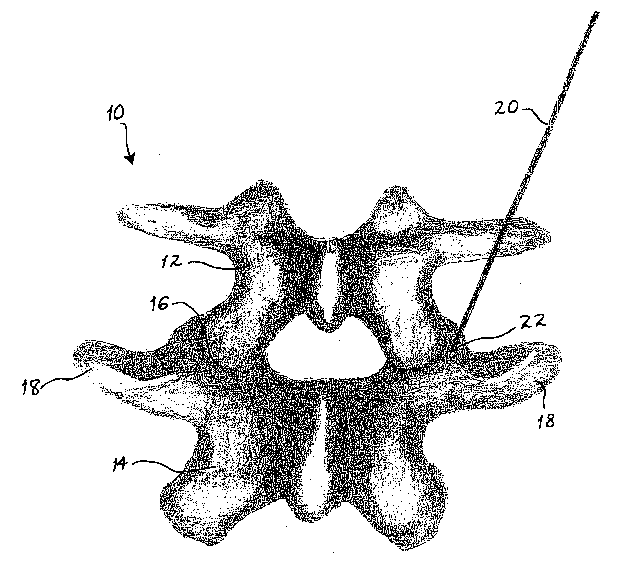
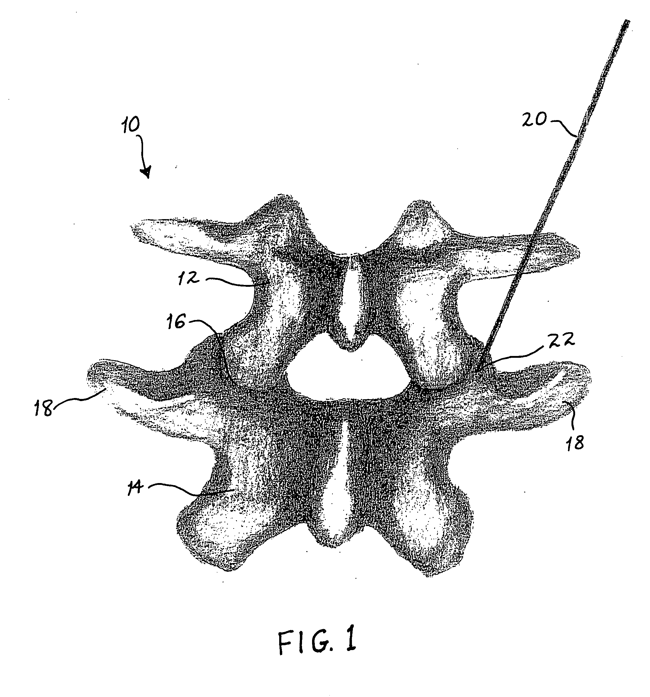
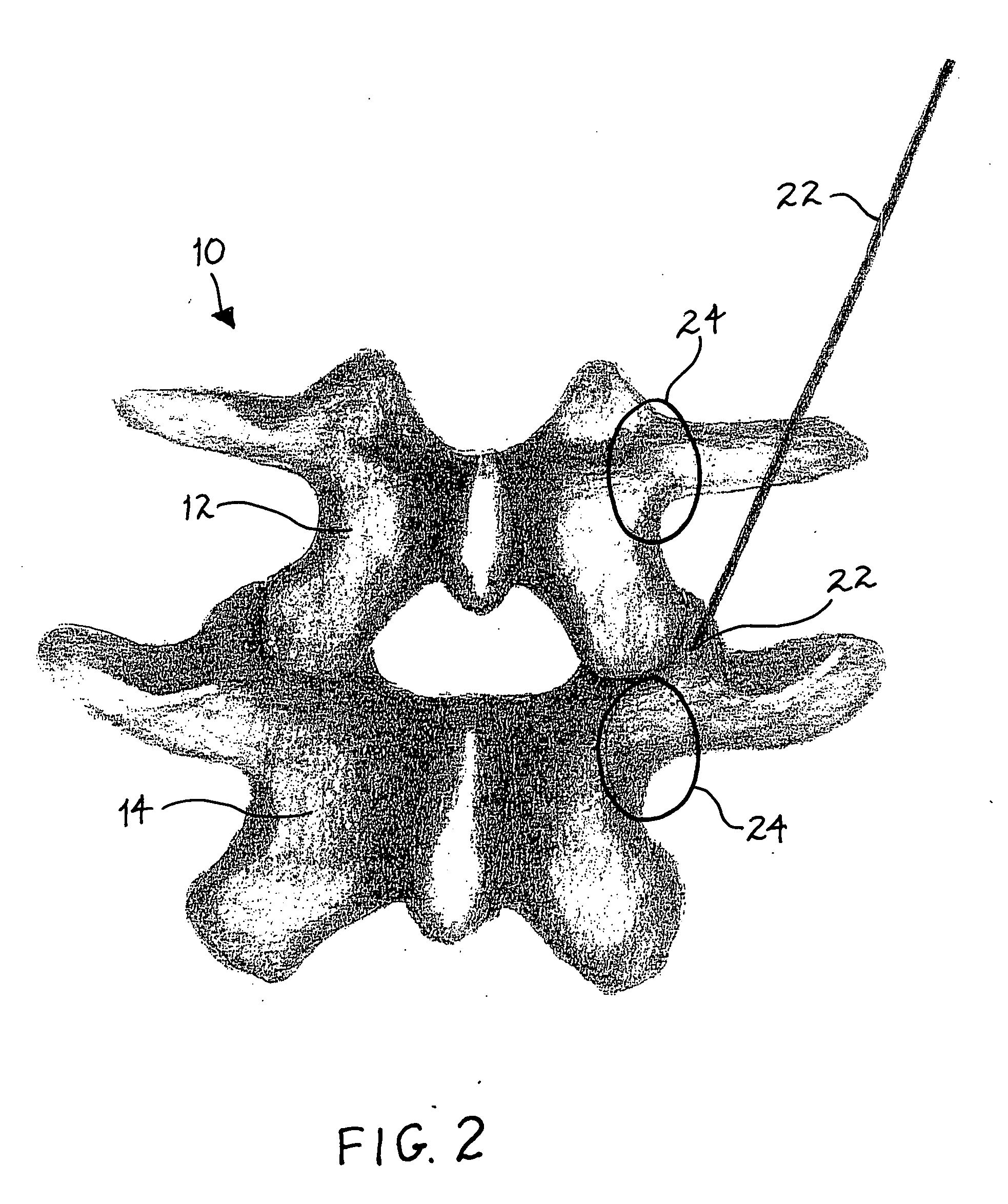
[0047] Referring now to the drawings, in which like reference numerals identify similar or identical elements throughout the many views, FIGS. 1-26 illustrate the minimally invasive fusion procedure of the present invention, while FIGS. 27 and 28 illustrate the novel bone graft capsules of the present invention, which facilitate the fusion procedure. The fusion procedure of the present invention can be carried out in many ways, but is preferably done through small incisions under fluoroscopy or other imaging procedure to view the surgical process. That is, the method of the present invention may be performed percutaneously, endoscopically, or even in a traditional “open” surgical procedure. Preferably, the method is performed percutaneously.

[0048] The fusion procedure preferably supplements a stabilization procedure, which, for example, provides pedicle screws and connecting rods between the pedicle screws. In such stabilization procedure, a series of dilators are passed over a gui...


PUM

PropertyMeasurementUnit
90° angleaaaaaaaaaa
bioabsorbableaaaaaaaaaa
viscousaaaaaaaaaa

Abstract

A method for performing a spinal fusion procedure between the transverse processes of two adjacent vertebra is disclosed. The method includes creating a vascularized bone flap at each of the transverse processes and introducing a bone graft delivery device to the site between the transverse processes and the vascularized bone grafts. A novel bone graft delivery system and device is also disclosed.

Description

CROSS-REFERENCE TO RELATED APPLICATION [0001] This application is based on, and claims priority to, U.S. Provisional Patent Application No. 60 / 549,695 filed on Mar. 3, 2004, the entire contents of which are hereby incorporated by reference.BACKGROUND [0002] 1. Field of the Invention [0003] The present invention relates to a minimally invasive method for performing spinal fusion procedures. In particular, the invention relates to a method for subcutaneously supplementing a stabilization procedure using pedicle screws and rods to stabilize adjacent vertebra, by utilizing a minimally-invasive-posterior-approach method of implanting bone growth material in the form of bone graft capsule to create a bone bridge between the adjacent vertebra. [0004] 2. Description of the Related Art [0005] Surgical treatment for spine disease may involve stabilization of the spine following disc removal and implantation of artificial discs. A common stabilization technique provides for screws to be secure...

Claims


Application Information

Patent Timeline
Patent Type & Authority Applications(United States)
IPC IPC(8): A61B17/56
CPCA61B17/14A61B17/148A61B2017/564A61F2/28A61F2/4455A61F2002/2817A61F2002/2835A61F2002/30062A61F2002/30242A61F2002/30462A61F2002/30588A61F2210/0004A61F2220/0075A61F2230/0071A61F2310/00023A61B17/707A61B17/144
Inventor BOEHM, FRANK H. JR.MELNICK, BENEDETTA DELORENZO
Owner BOEHM FR H JR
Why Eureka
Try Eureka