Methods and apparatus for non-invasively treating atrial fibrillation using high intensity focused ultrasound

Inactive Publication Date: 2005-10-13
TERUMO KK
View PDF99 Cites 24 Cited by
  • Summary
  • Abstract
  • Description
  • Claims
  • Application Information

AI Technical Summary

Benefits of technology

[0018] It is another object of the present invention to provide methods and apparatus for treating PFO by applying energy from outside the body or from neighboring organs, such as the esophagus, that are easily accessible.
[0019] These and other objects of the present invention are accomplished by providing methods and apparatus that enable a physician to image tissue within

Problems solved by technology

These, however are invasive devices whic

Method used


Image

  • Methods and apparatus for non-invasively treating atrial fibrillation using high intensity focused ultrasound
  • Methods and apparatus for non-invasively treating atrial fibrillation using high intensity focused ultrasound
  • Methods and apparatus for non-invasively treating atrial fibrillation using high intensity focused ultrasound

Examples


Embodiment Construction

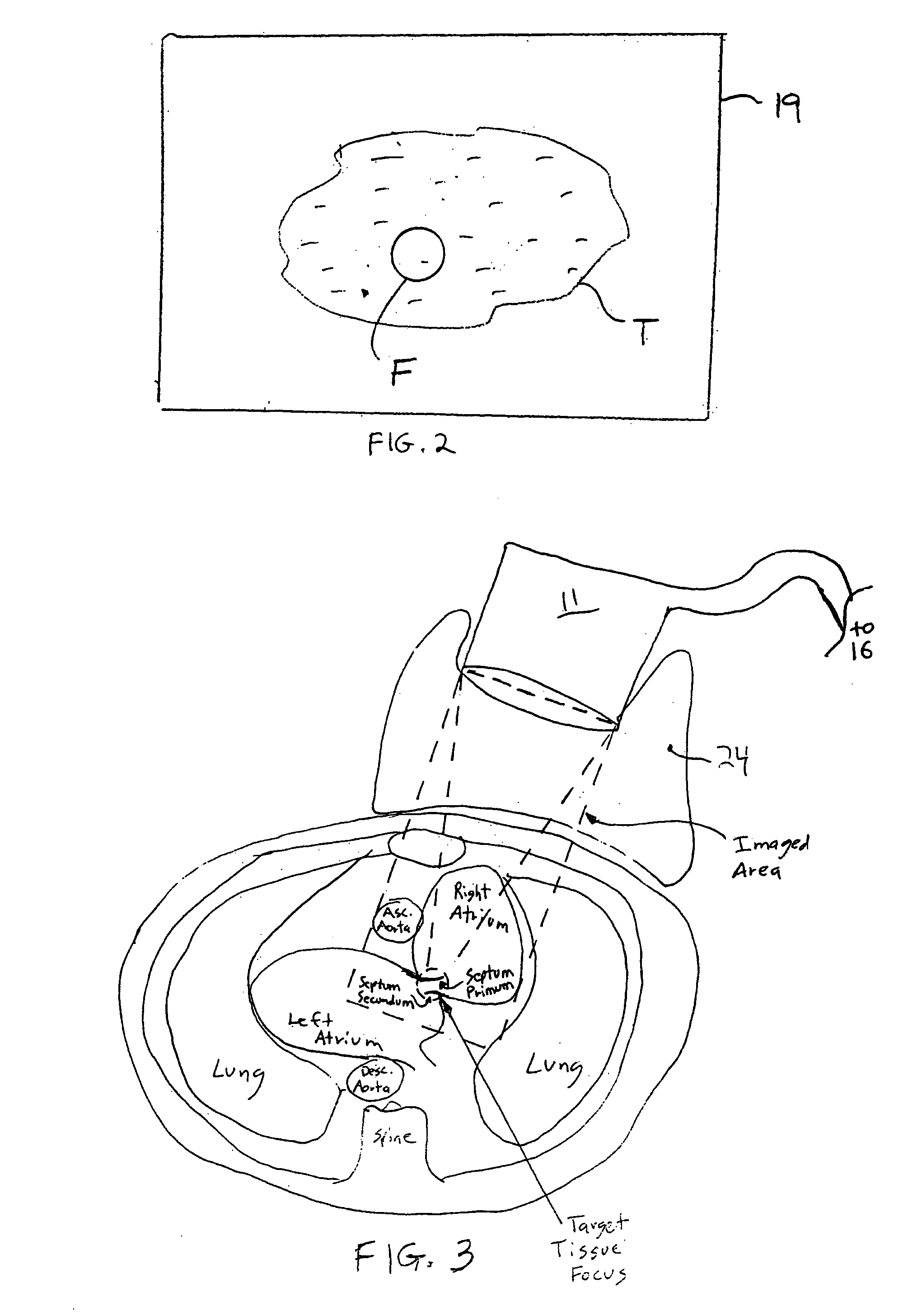
[0025] The present invention is directed to methods and apparatus for creating lesions in the walls of the heart or for ablating or welding the tissue of a PFO in a non-invasive manner using high intensity focused ultrasound (HIFU). Previously-known HIFU systems, such as those being developed by Epicor Medical or Transurgical, require close approximation of the HIFU device to the target tissue. These systems are not adapted for PFO closure. The methods and apparatus of the present invention overcome this drawback by providing systems that enable the creation of lesions in the heart wall from a greater distance.

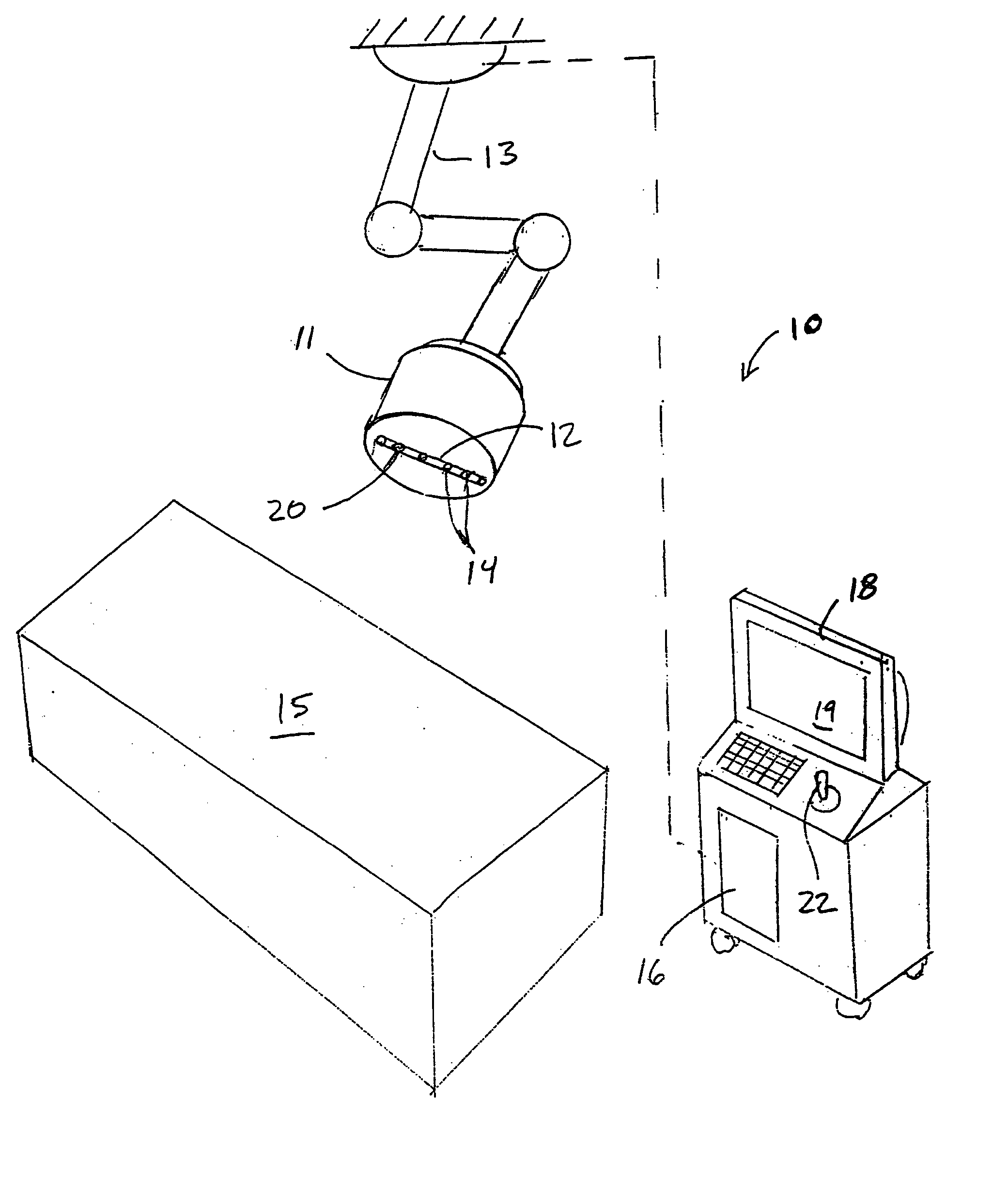
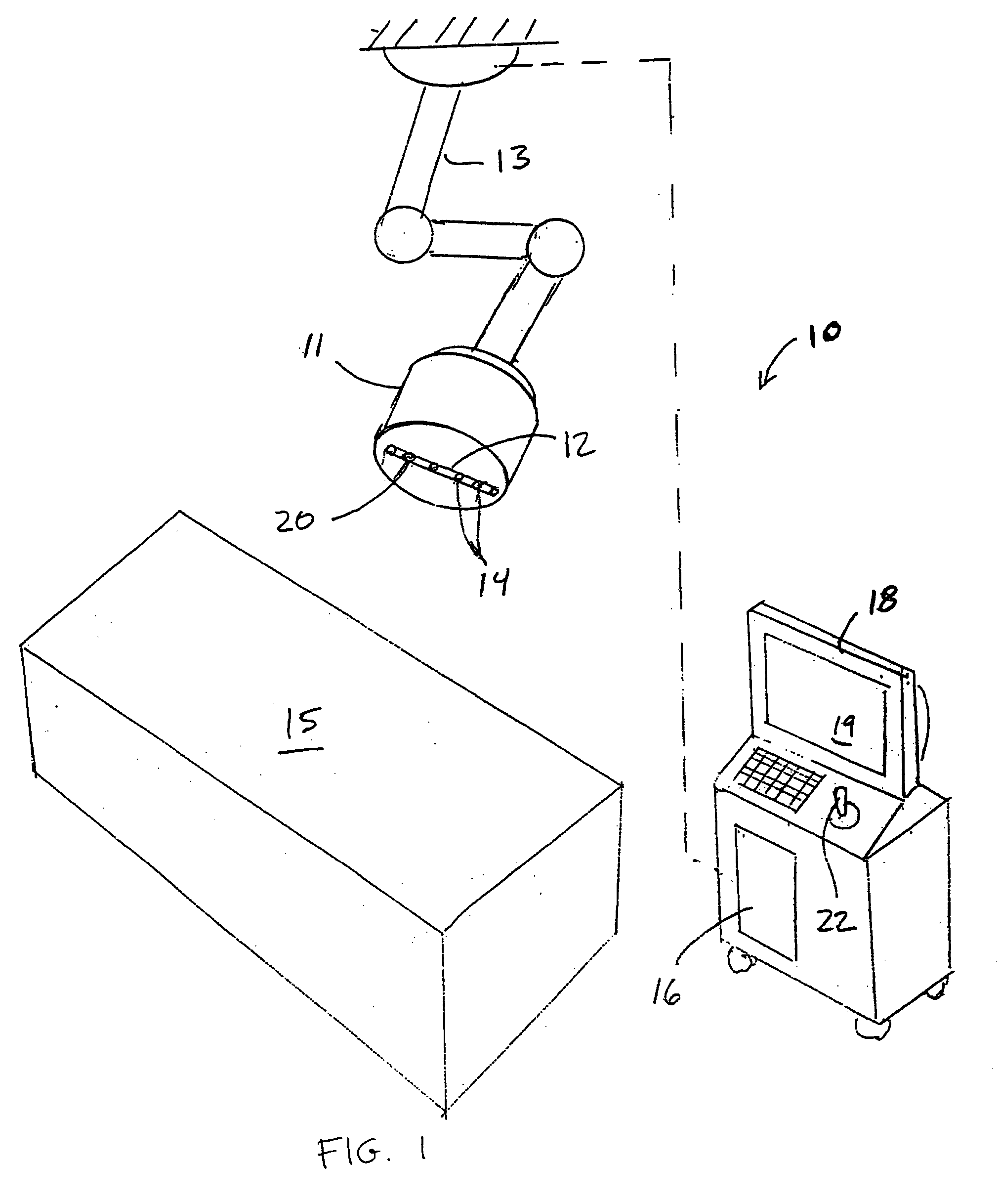
[0026] Referring to FIG. 1, apparatus constructed in accordance with the principles of the present invention is described. System 10 comprises head 11 housing ultrasound imaging system 12 and high intensity focused ultrasound energy (“HIFU”) system 14. Ultrasound imaging system 12 and HIFU system 14 may have in common all or just a subset of the transducers and related compon...


PUM


Abstract

Methods and apparatus are provided for non-invasively treating patent foramen ovale using an ultrasound imaging system and a high intensity focused ultrasound system to selectively target high intensity ultrasound energy on either or both of a patient's septum primum or septum secundum.

Description

REFERENCE TO RELATED APPLICATIONS [0001] This application claims priority from and is a Continuation-In-Part of U.S. provisional patent application Ser. No. 60 / 477,532, filed Jun. 10, 2003.FIELD OF THE INVENTION [0002] The present invention relates to methods and apparatus for treatment of patent foramen ovale, and more particularly, to the use of high intensity focused ultrasound non-invasively to treat patent foramen ovale. BACKGROUND OF THE INVENTION [0003] Up until the 1980s, there was dramatic growth in the creation of new surgical methods for treating a wide variety of previously untreated conditions. Over the past twenty years there has been a clear trend towards the invention of devices and methods that enable less-invasive treatment of such diseases, moving from invasive surgery, and then to less-invasive surgery, and to interventional techniques. Ultimately, it is desirable to move to totally non-invasive therapies. [0004] Patent foramen ovale (PFO) is characterized by a p...

Claims


Application Information

Patent Timeline
IPC IPC(8): A61BA61B17/00A61B18/04A61B18/18A61N7/00A61N7/02
CPCA61B8/0883A61B8/12A61B8/4488A61N2007/0078A61B2019/5278A61N7/02A61B2017/00243A61B2018/00351A61B2090/3782
Inventor GIFFORD, HANSON S.DEEM, MARK
Owner TERUMO KK
Why Eureka
Try Eureka