Apparatus and method for forming 3D ultrasound image

a 3d ultrasound and image technology, applied in the field of 3d ultrasound diagnostic system, can solve the problems of incongruity of roi box size and roi shape, inability to accurately display the 3d ultrasound image of the target object, and difficulty in clearly displaying the shape of the fetus with the 3d ultrasound image, so as to reduce errors and reduce time consumed

Active Publication Date: 2005-10-27
MEDISON CO LTD
View PDF35 Cites 56 Cited by
  • Summary
  • Abstract
  • Description
  • Claims
  • Application Information

AI Technical Summary

Benefits of technology

[0010] Therefore, it is an objective of the present invention to provide an apparatus and a method for forming an accurate 3D ultrasound image of a target object while reducing errors, which may be generated in a rendering process of a volume data and a contour detection process of the target object. ...

Problems solved by technology

However, since the volume data includes mixed data of uterus tissue, adipose tissue, amniotic fluid, floating matters and the fetus, if the rendering process is directly applied to the volume data, it is difficult to clearly display the shape of the fetus with the 3D ultrasound image.
That is, the size of the ROI box is not consistent according to the user generating the ROI box.
As such, there is often a problem since a desired 3D ultrasound image of the target object cannot be ac...

Method used


Image

  • Apparatus and method for forming 3D ultrasound image
  • Apparatus and method for forming 3D ultrasound image
  • Apparatus and method for forming 3D ultrasound image

Examples


Embodiment Construction

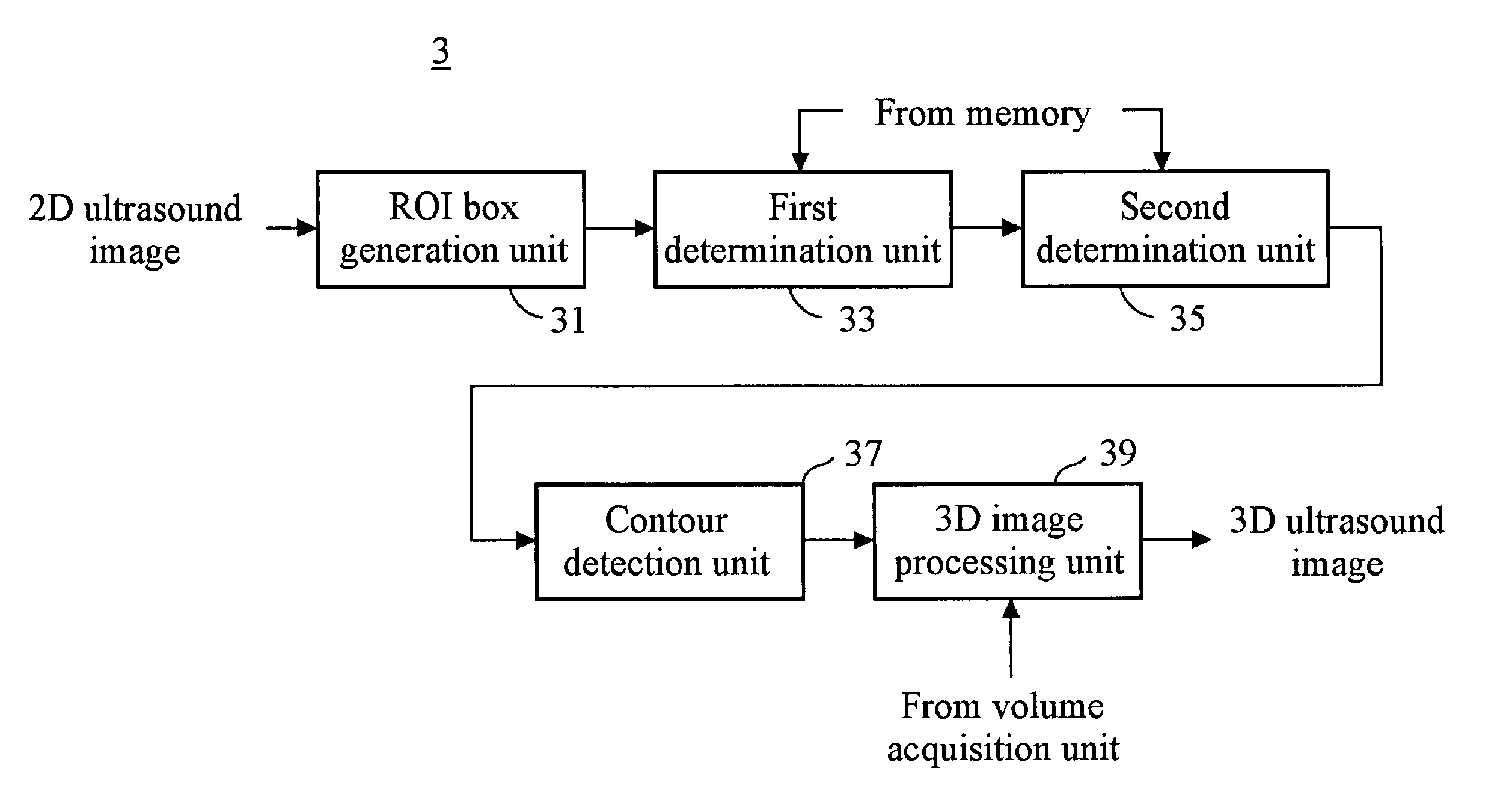
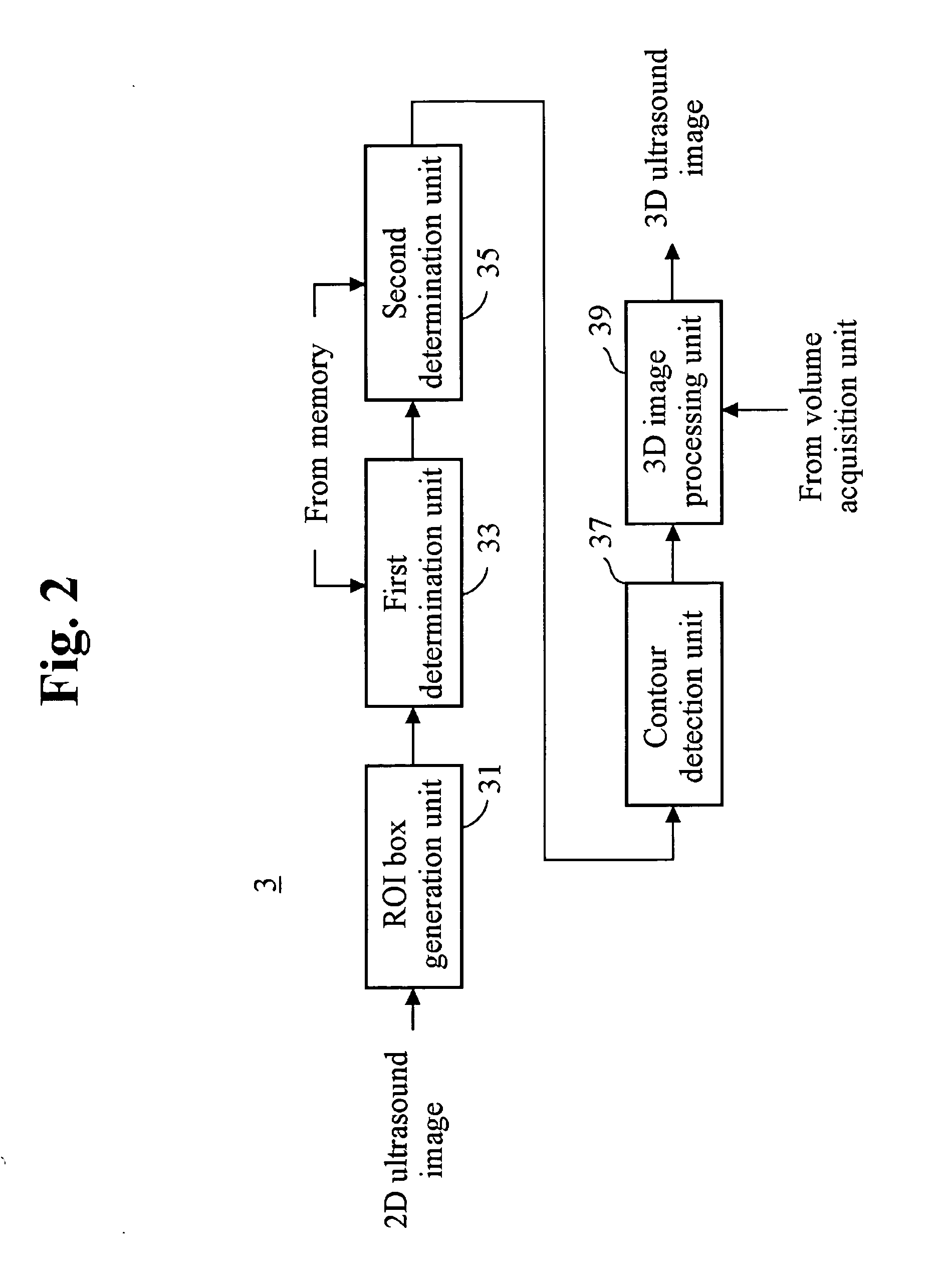
[0024]FIG. 2 is a schematic block diagram showing a 3-dimensional (3D) ultrasound image forming device 3 in a 3D ultrasound diagnostic device constructed in accordance with the preferred embodiment of the invention. The 3D ultrasound image forming device 3 includes a region of interest (ROI) box generation unit 31, a first determination unit 33, a second determination unit 35, a contour detection unit 37 and a 3D image processing unit 39. As the 3D key of a control panel (not shown) mounted in the 3D ultrasound image forming device 3 is activated, the 3D ultrasound image forming device 3 starts to operate.

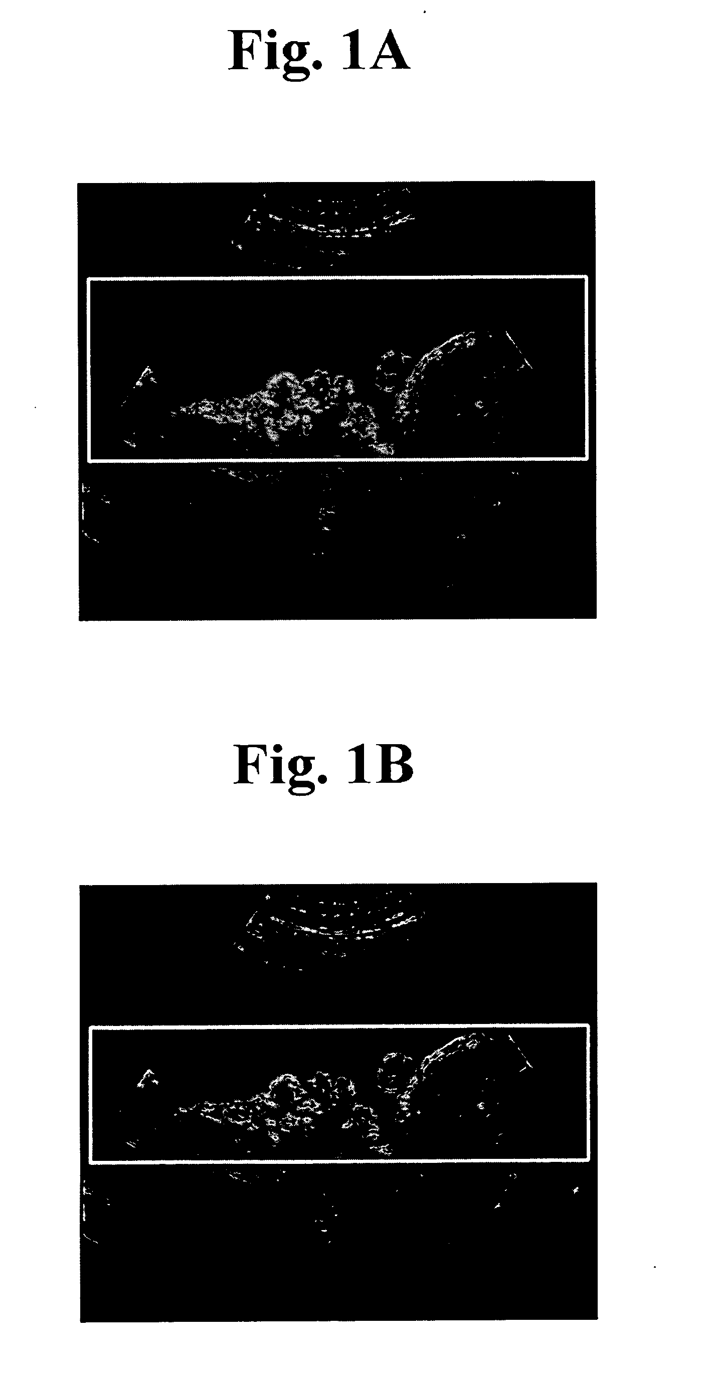
[0025] If a user activates the 3D key, then one of the 2D ultrasound images, which are acquired through a probe and a beam former in the 3D ultrasound diagnostic system, is displayed with a brightness mode (B-mode) on a display device (not shown). In accordance with the present invention, the 2D ultrasound image displayed on the display device is a 2D ultrasound image representing...


PUM


Abstract

The present invention relates to a 3D ultrasound diagnostic forming a 3D ultrasound image only with volume data exiting within contour by automatically detecting the contour of a target object, comprising: a first unit for generating a region of interest (ROI) box on a 2D ultrasound image; a second unit for detecting a contour of a target object in the ROI box; and a third unit for forming a 3D ultrasound image by rendering volume data existing in the detected contour.

Description

FIELD OF THE INVENTION [0001] The present invention generally relates to a 3-dimensional (3D) ultrasound diagnostic system, and more particularly to an apparatus and a method for automatically detecting a contour from a 2-dimensional (2D) ultrasound image of a target object and forming a 3D ultrasound image with volume data within the contour. BACKGROUND OF THE INVENTION [0002] A 3-dimensional (3D) ultrasound diagnostic system is a medical equipment for providing clinical information such as spatial information, anatomical information and the like, which cannot be provided from a conventional 2-dimensional image. The 3D ultrasound diagnostic system acquires volume data from signals received from a target object through a probe, and performs a scan conversion process for the acquired volume data. A 3D ultrasound image of the target object is displayed on a display device such as a monitor, a screen or the like by performing a rendering process upon images obtained from the scan-conve...

Claims


Application Information

Patent Timeline
IPC IPC(8): A61B8/00G01S7/539G01S15/89G06T15/08
CPCA61B8/00A61B8/483A61B8/467A61B8/461A61B8/469Y10S128/916E02D29/0275E02D2250/0007E02D2250/0023
Inventor SHIM, JAEYOONKWAK, BYOUNG JOOKIM, NAM CHULKIM, SANG HYUN
Owner MEDISON CO LTD
Why Eureka
Try Eureka