Radiographic image capturing apparatus

a technology of radiographic images and capturing apparatuses, applied in the field of radiographic image capturing apparatuses, can solve the problems of difficult to find out such timing, increase in medical cost, wasteful medical cost, etc., and achieve the effect of simple operation

Inactive Publication Date: 2005-11-03
CANON KK
View PDF8 Cites 15 Cited by
  • Summary
  • Abstract
  • Description
  • Claims
  • Application Information

AI Technical Summary

Benefits of technology

[0008] The present invention has been made in consideration of the aforementioned problems, and

Problems solved by technology

Furthermore, the diagnostic workup may often determine that the initial diagnosis is a false diagnosis, and no disease is found.
Hence, wasteful medical cost is spent, resulting in part of the reason of medical cost i

Method used


Image

  • Radiographic image capturing apparatus
  • Radiographic image capturing apparatus
  • Radiographic image capturing apparatus

Examples


first embodiment

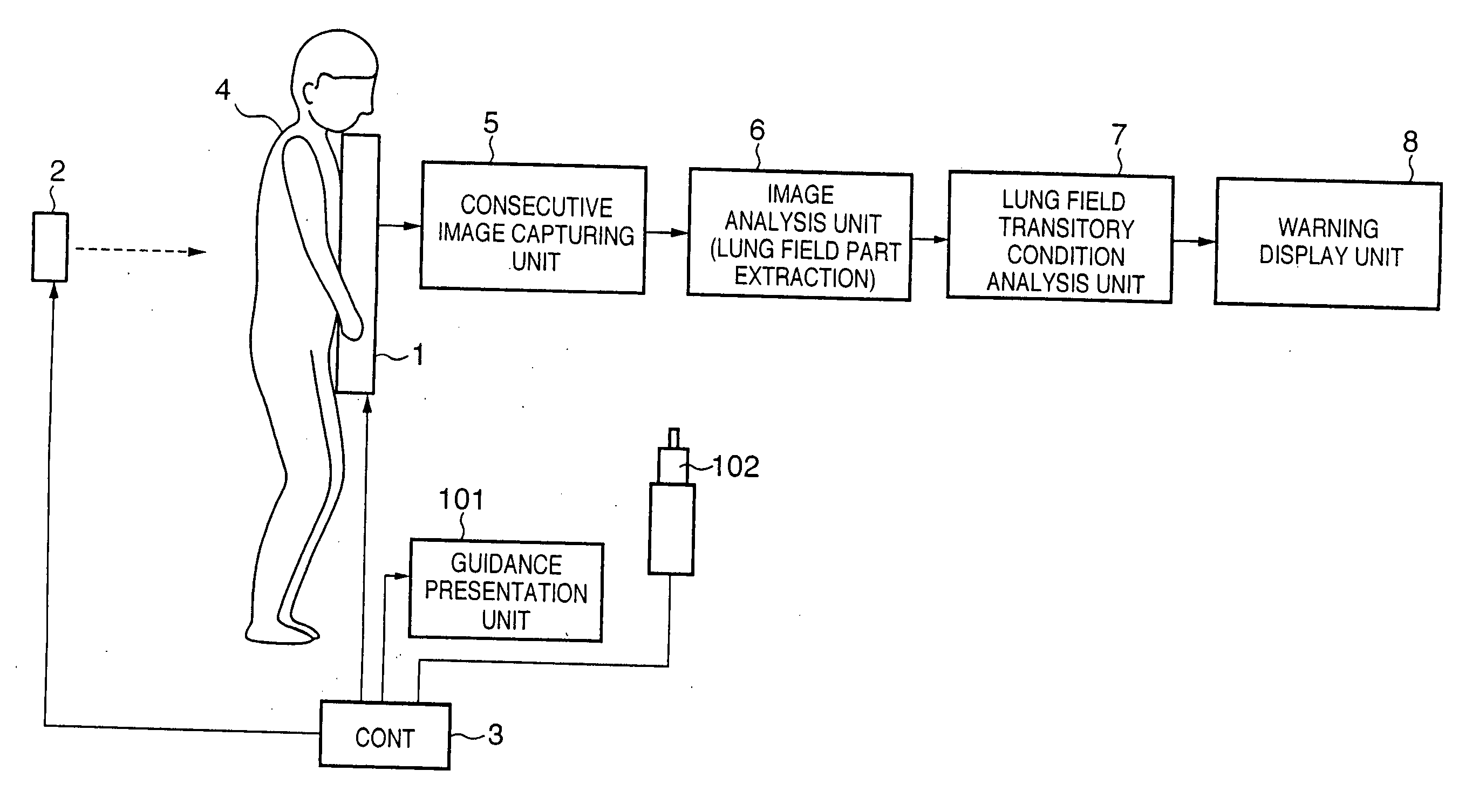
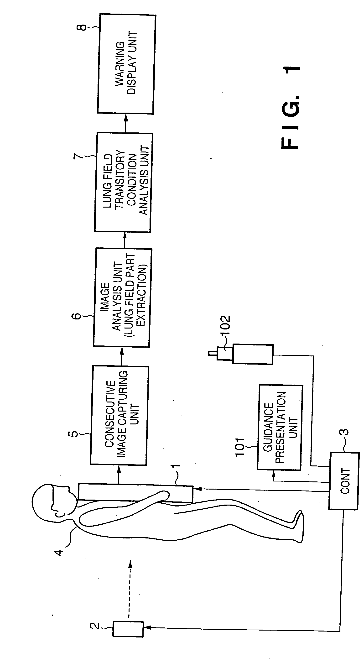
[0021] In the first embodiment, captured respiratory behavior images are analyzed in real time (parallel to moving image capturing) to detect the transitory condition of the size of a lung field part, and it is checked based on the transitory condition if appropriate respiratory behavior images are obtained. That is, a plurality of images are analyzed while they are captured, and the analysis result (advisability of images) is quickly presented after completion of imaging. If it is determined that images are inappropriate, imaging is prompted again. In the following embodiments, an X-ray image is used as a radiographic image.

[0022]FIG. 1 is a block diagram showing the arrangement of a respiratory behavior imaging apparatus in the first embodiment. Referring to FIG. 1, an X-ray image sensor 1 detects and images the intensity distribution of X-rays which are irradiated from an X-ray generator 2 and reach that sensor. The X-ray image sensor 1 comprises an FPD, and can consecutively cap...

second embodiment

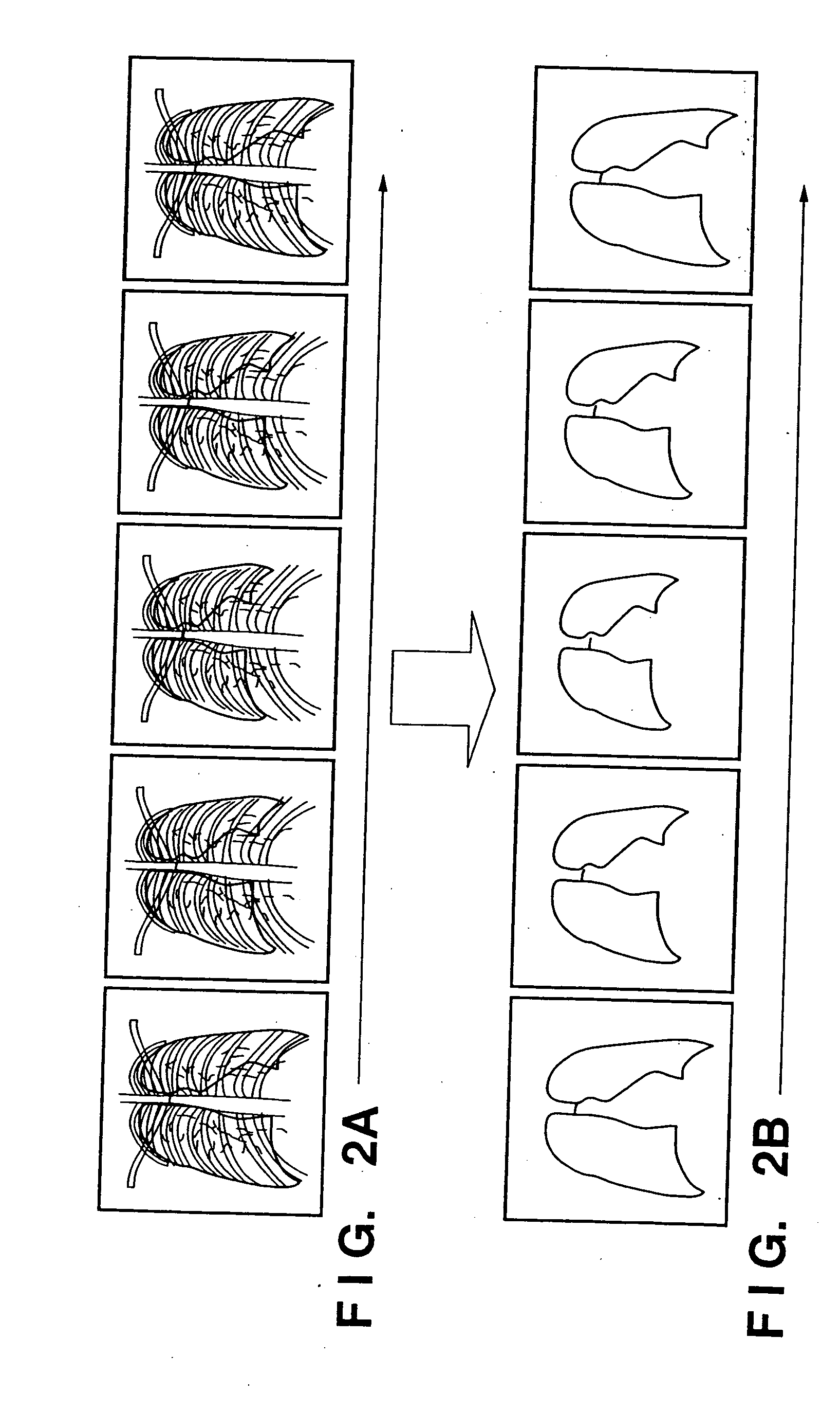
[0036]FIGS. 5A to 5D are timing charts indicating the operation timings of the respiratory behavior imaging process according to the The lung field transitory condition analysis unit 7 outputs and analyzes the fluctuation of the lung field lengths, as shown in FIG. 5A. Note that detection of the fluctuation of the lung field lengths is as has been explained above with reference to FIGS. 2 and 3. Note that the advisability of respiratory actions can be determined by various methods. For example, in this embodiment, it is determined that an appropriate respiratory condition is obtained when a time interval (T1) between the neighboring peaks on the maximum side of the lung field lengths, a time interval (T2) between the neighboring maximum and minimum peaks, and a fluctuation (L) in the graph shown in FIG. 5A fall within a predetermined range for a predetermined period.

[0037] The Ready display unit 9 makes an imaging ready display at a timing 11 at which it can be determined that the ...


PUM


Abstract

A radiographic image capturing apparatus captures consecutive radiographic images on the basis of the radiation intensity distribution transmitted through a subject, and extracts lung field parts from the captured consecutive radiographic images. A fluctuation condition is detected from the extracted lung field parts, and whether or not the respiratory condition of the subject is suited to radiograph a respiratory behavior is determined on the basis of the detected fluctuation condition.

Description

FIELD OF THE INVENTION [0001] The present invention relates to imaging using radiation in the medical field. BACKGROUND OF THE INVENTION [0002] A technique for imaging a transmission intensity distribution using the material transmission performance of radiation represented by X-rays is fundamental of development in modern medical techniques. Since discovery of X-rays, imaging of the intensity distribution of radiation adopts a method in which an X-ray intensity distribution is converted into visible light by a phosphor, a latent image is formed by using a silver halide film, and the latent image is developed. In recent years, a method of using a so-called imaging plate is generally used. This method uses a photostimulable phosphor upon converting an X-ray image into a digital image, and reads out a latent image as an accumulated energy distribution on the photostimulable phosphor by exciting it by a laser beam, thus capturing a digital image. Furthermore, with the advance of the se...

Claims


Application Information

Patent Timeline
IPC IPC(8): G06K9/00A61B6/00G06T7/00
CPCA61B6/4233A61B6/50G06T2207/30061G06T7/0012A61B6/541
Inventor INOUE, HITOSHI
Owner CANON KK
Why Eureka
Try Eureka