System, apparatus, and method for supporting insertion of endoscope

a technology of endoscope and endoscope body, which is applied in the field of system, apparatus and method for supporting the insertion of endoscope, can solve the problems of difficult to confirm and correct the insertion direction of bronchoscop

Inactive Publication Date: 2005-11-24
OLYMPUS CORP
View PDF7 Cites 57 Cited by
  • Summary
  • Abstract
  • Description
  • Claims
  • Application Information

AI Technical Summary

Problems solved by technology

With respect to a multi-branching tubular organ, such as a bronchus dividing into smaller bronchi and subsequently bronchioles, if the whole bronchial tree can be displayed on a display screen, it is difficult to confirm and correct the insertion direction of a bronchoscope.

Method used


Image

  • System, apparatus, and method for supporting insertion of endoscope
  • System, apparatus, and method for supporting insertion of endoscope
  • System, apparatus, and method for supporting insertion of endoscope

Examples


Embodiment Construction

[0026] Embodiments of the present invention will now be described below with reference to the drawings.

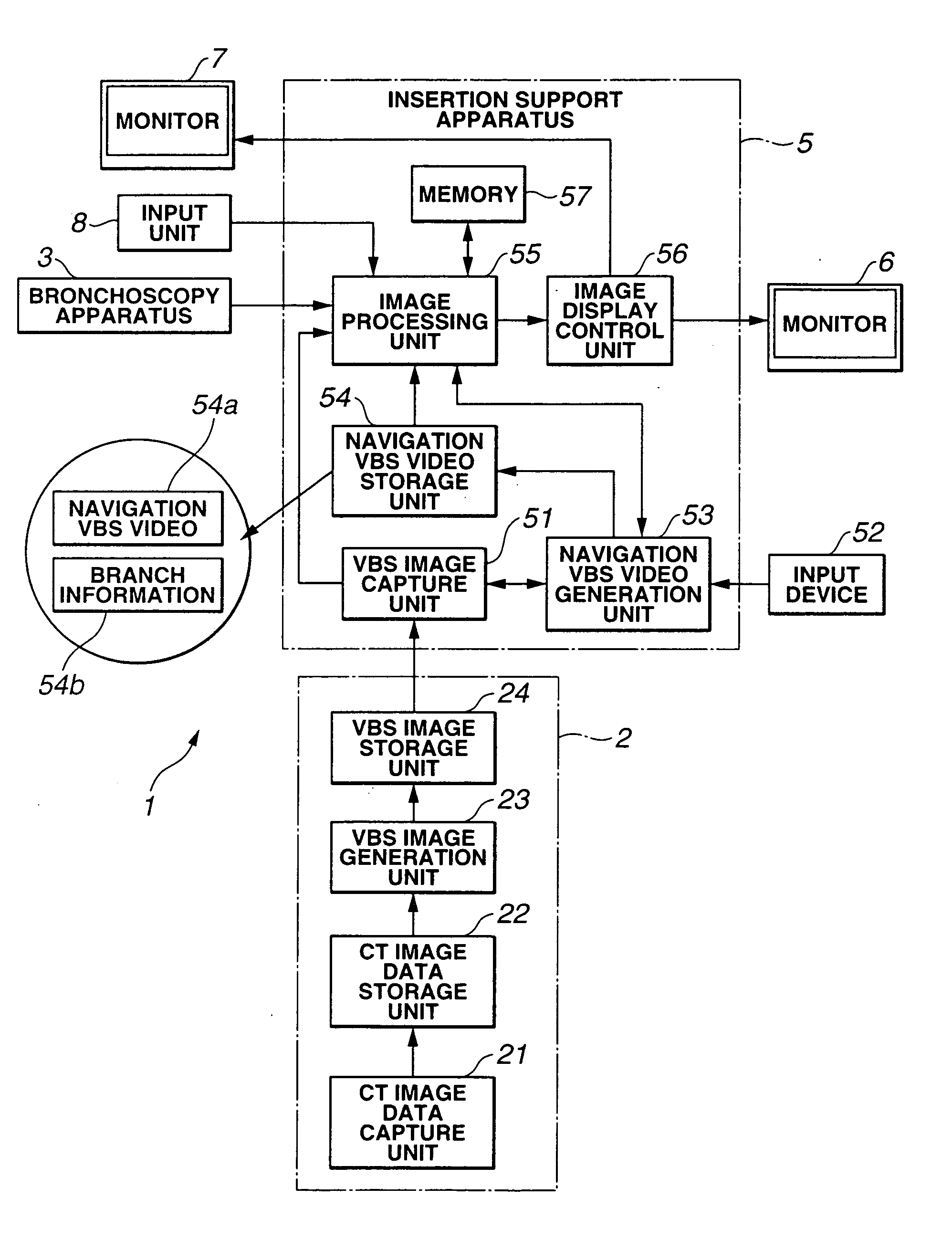
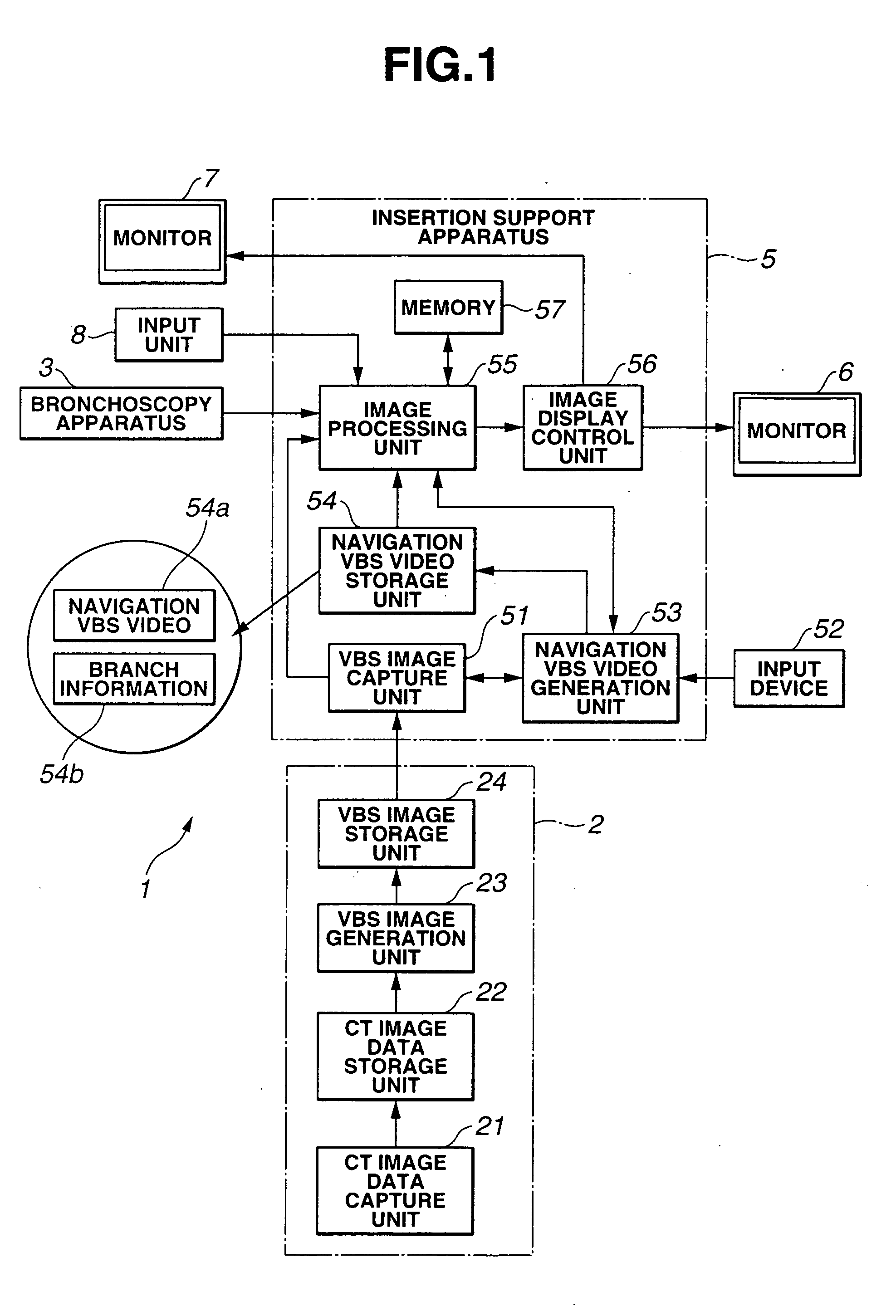
[0027]FIG. 1 shows a system 1 for supporting the insertion of an endoscope (bronchoscope) into a bronchus according to an embodiment of the present invention. The system 1 includes a VBS image generation apparatus 2 for generating a virtual endoscopic image of inside of bronchus according to a virtual bronchoscopy system (hereinafter, referred to as a VBS image), a bronchoscopy apparatus 3, and an insertion support apparatus 5. The VBS image generation apparatus 2 generates a VBS image based on CT image data. The insertion support apparatus 5 combines an endoscopic image (hereinafter, referred to as a live image) captured by the bronchoscopy apparatus 3 with the VBS image obtained by the VBS image generation apparatus 2 and displays the combined image in monitors 6 and 7 so as to support the insertion of the bronchoscopy apparatus 3 into a bronchus.

[0028] The bronchoscopy apparat...


PUM


Abstract

According to the present invention, to guide an endoscope to a target part in vivo with reliability on the basis of guide images corresponding to actual branch points, an endoscope insertion support system includes a VBS image capture unit for capturing VBS images stored in a VBS image storage unit, a navigation VBS video generation unit for generating a navigation VBS video serving as a series of navigation moving pictures to support the insertion of a bronchoscope into a bronchus on the basis of patient information entered from an input device, a navigation VBS video storage unit for storing the navigation VBS video, an image processing unit for performing various image processing operations, and a memory for temporarily storing registered navigation VBS frame images while the navigation VBS video is being generated.

Description

[0001] This application claims benefit of Japanese Patent Application No. 2004-24834 filed in Japan on Jan. 30, 2004, the contents of which are incorporated by this reference. BACKGROUND OF THE INVENTION [0002] 1. Field of the Invention [0003] The present invention relates to a system, apparatus, and method for supporting the insertion of an endoscope. [0004] 2. Description of the Related Art [0005] In the medical diagnostic field, diagnoses based on images are coming into wide use. As one of the diagnoses, tomograms of a part of a subject are captured using an x-ray computed tomography (CT) system to obtain three-dimensional (3D) image data of the part. The subject is diagnosed on the basis of the 3D image data of the part. In the CT system, an x-ray generation unit and an x-ray detection unit are continuously rotated around a subject at the same time as the subject is continuously moved along the body axis, thus performing a spiral continuous scan (helical scan) to the subject in ...

Claims


Application Information

Patent Timeline
Patent Type & Authority Applications(United States)
IPC IPC(8): G02B23/24A61B1/00A61B1/04A61B19/00G06T1/00
CPCA61B1/00009A61B19/5225A61B19/50A61B19/20A61B90/10A61B34/10A61B90/37
Inventor AKIMOTO, SHUNYAONISHI, JUNICHI
Owner OLYMPUS CORP
Why Eureka
Try Eureka