

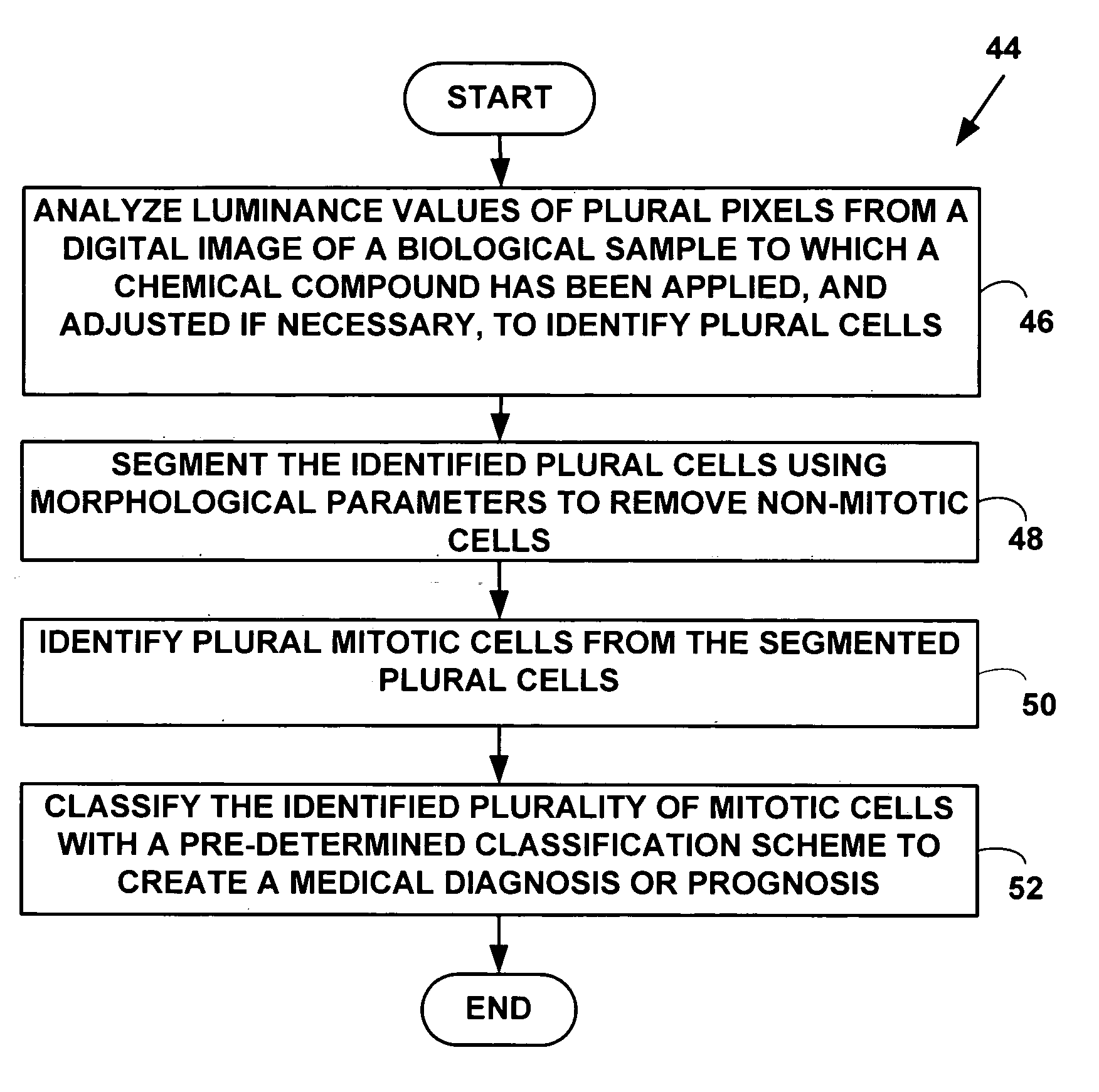
Method and system for morphology based mitosis identification and classification of digital images
a technology of morphology and mitosis, applied in the field of digital image processing, can solve the problems of complex and involved determination of mitotic count, insufficient information for referring medical clinicians to make decisions about patient prognosis and treatment, and many pitfalls, so as to improve the prognosis and selection
- Summary
- Abstract
- Description
- Claims
- Application Information
AI Technical Summary
Benefits of technology
Problems solved by technology
Method used
Image

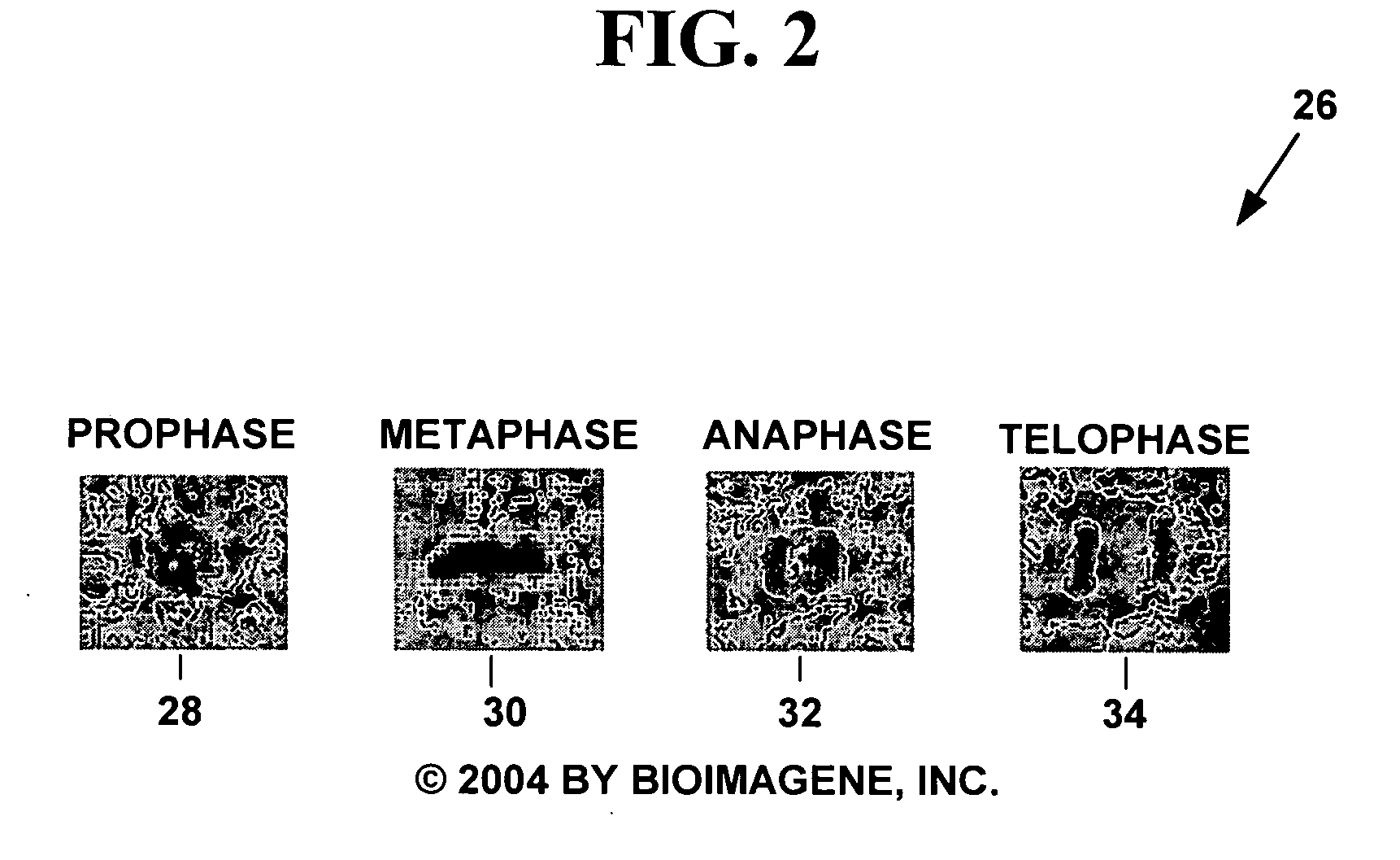
Examples
Embodiment Construction
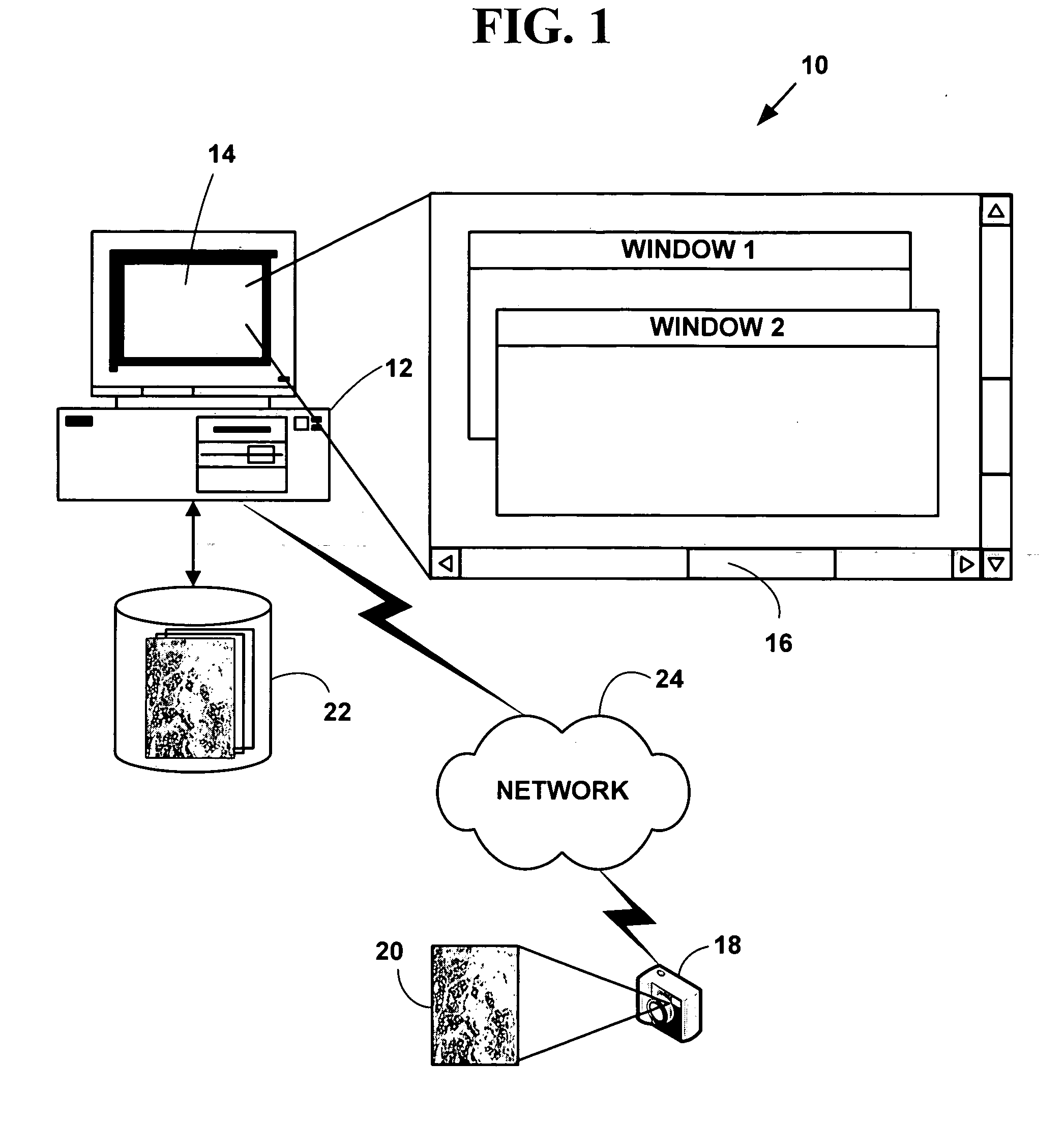
Exemplary Mitosis Identification and Classification System
[0069]FIG. 1 is a block diagram illustrating an exemplary automated digital image based mitosis detection and classification system 10. The exemplary system 10 includes one or more computers 12 with a computer display 14 (one of which is illustrated). The computer display 14 presents a windowed graphical user interface (“GUI”) 16 with multiple windows to a user. The system 10 may optionally include a microscope or other magnifying device (not illustrated in FIG. 1). The system 10 further includes a digital camera 18 (or analog camera) used to provide plural digital images 20 in various digital images or digital data formats. One or more databases 22 (one or which is illustrated) include biological sample information in various digital images or digital data formats. The one or more database 22 may also include raw and processed digital images and may further include knowledge databases created from automated analysis of the...
PUM
| Property | Measurement | Unit |
|---|---|---|
| diameter | aaaaa | aaaaa |
| field surface area | aaaaa | aaaaa |
| threshold value | aaaaa | aaaaa |
Abstract
Description
Claims
Application Information