Lesion boundary detection

a lesion and boundary detection technology, applied in the field of lesion boundary detection, can solve the problems of difficult to detect the extent of a lung nodule attached to the pleura, difficult to separate a nodule, difficult to detect the boundary between a polyp and the colon wall,

a lesion and boundary detection technology, applied in the field of lesion boundary detection, can solve the problems of difficult to detect the extent of a lung nodule attached to the pleura, difficult to separate a nodule, difficult to detect the boundary between a polyp and the colon wall,

US20050286750A1Inactive Publication Date: 2005-12-29SAMSUNG ELECTRONICS CO LTD

Image

  • Lesion boundary detection
  • Lesion boundary detection
  • Lesion boundary detection

Examples


first embodiment

[0101] In a first embodiment, an approximation may be made that the nodule N is not attached to or proximate to any features other than the pleura P. The result of the segmentation step may be used to identify all of the foreground region beyond the line joining the junction points as forming part of the nodule N, as shown in FIG. 15.

second embodiment

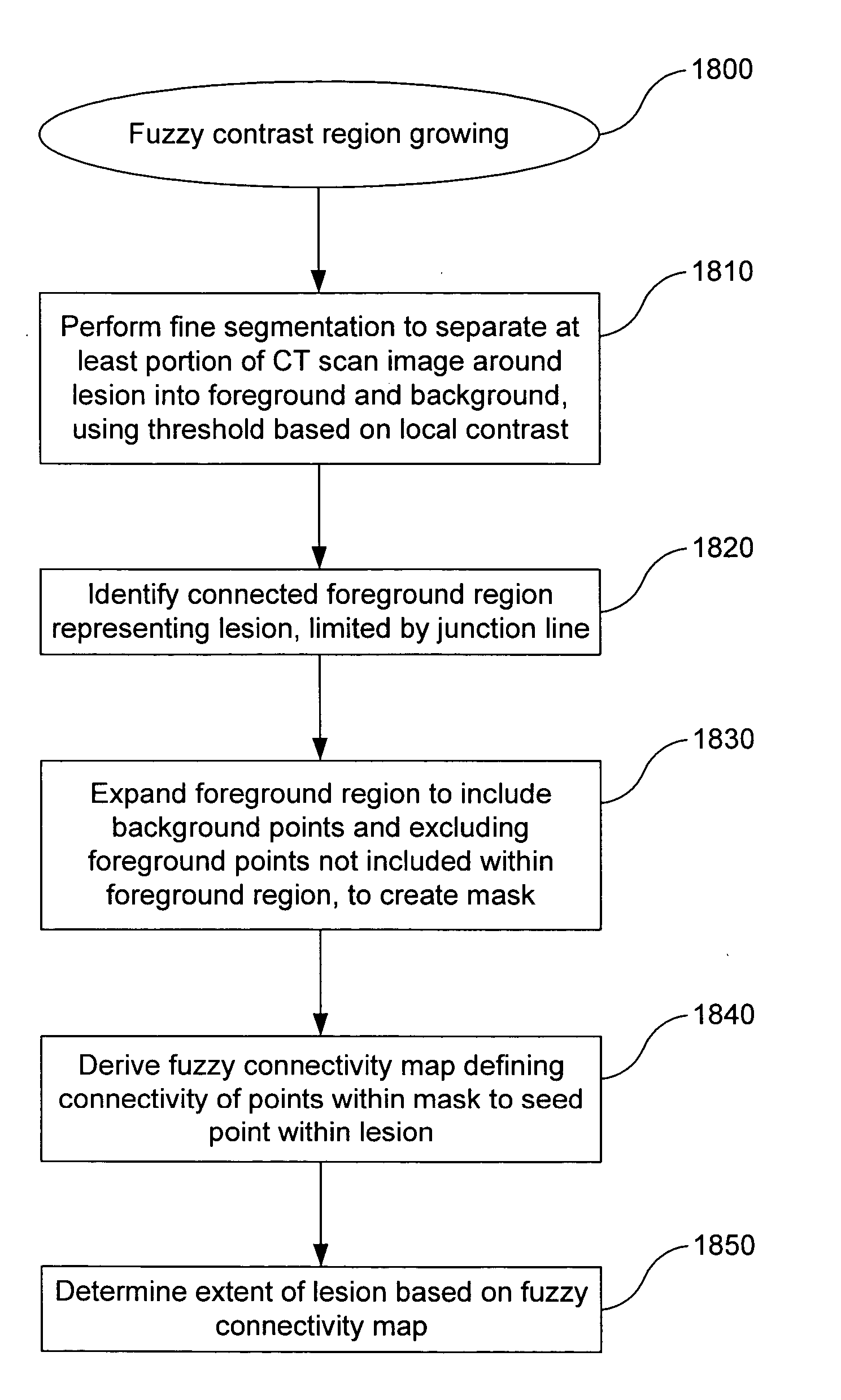
[0102] In a second embodiment, and referring to FIG. 18, a fuzzy contrast region growing scheme 1800 may be used to identify the boundary of the nodule N. A rough estimate of the extent of the nodule N may be obtained by means of a local threshold-based segmentation process at block 1810. A local mask may be used to derive a threshold intensity value for each point. The threshold typically is sensitive to the local contrast and usually can distinguish low contrast nodules from their background. The nodule area N acquired by fine segmentation may include ‘holes’, i.e. background pixels surrounded by foreground pixels. The holes can occur based on the sensitivity of the fine segmentation to small local differences in contrast. A hole-filling algorithm may be used to fill such holes. For instance, the hole-filling algorithm may convert the holes to foreground pixels. Any suitable hole-filling algorithm may be used.

[0103] The pixel having the highest intensity within the nodule area N i...


PUM


Abstract

A method of detecting a junction between a lesion and a wall in a CT scan image may include determining the boundary (B) of the wall to an internal space (L), identifying critical points (c1, c2) along the boundary, and selecting one critical point at either side of the lesion as a junction point between the wall and the lesion. The critical points may be points of maximum local curvature and / or points of transition between straight and curved sections of the boundary. The critical points may be selected by receiving first and second seed points (p1, p2) at either side of the lesion, moving the seed points to the boundary if they are not already located on the boundary, and finding the closest critical points to the seed points. The seed points may be determined by displacing the determined junction points (j1, j2) from an adjacent slice of the image into the current slice.

Description

BACKGROUND OF THE INVENTION [0001] 1. Field of the Invention [0002] The present invention relates to a method of detecting the junction of a lesion and a wall, such as a lung nodule attached to pleura or a polyp attached to a colon wall, in an image scanned from a human or animal body, particularly but not exclusively in a computed tomography (CT) image. The invention encompasses software and apparatus for carrying out the method. [0003] 2. Background Art [0004] Detection of suspicious lesions in the early stages of cancer can be considered the most effective way to improve survival. Lung nodule detection and polyp detection are some of the more challenging tasks in medical imaging. [0005] Computer-assisted techniques have been proposed to identify regions of interest containing a nodule in a CT scan image, to segment the nodule from surrounding objects such as blood vessels or the lung wall, to calculate physical characteristics of the nodule, and / or to provide an automated diagnos...

Claims


Application Information

Patent Timeline
29 Dec 2005
Publication
US20050286750A1
IPC
A61B5/053; A61B6/03; G06F19/00; G06K9/00; G06T5/00
CPC
G06K9/342; G06K9/4638; G06K2209/05; G06T7/0081; G06T2207/30064; G06T2207/10081; G06T2207/20012; G06T2207/20104
Inventors
DEHMESHKI, JAMSHID