Computer-aided diagnostic apparatus

a computer and diagnostic technology, applied in the direction of magnetic variable regulation, instruments, image enhancement, etc., can solve the problems of difficult detection of tumors located at the apex of the lung, difficult to find tumors at the back of the heart, and difficult to obtain contrast, so as to facilitate double checking of the candidate for the sick portion

Active Publication Date: 2006-03-09
TOSHIBA MEDICAL SYST CORP
View PDF18 Cites 36 Cited by
  • Summary
  • Abstract
  • Description
  • Claims
  • Application Information

AI Technical Summary

Benefits of technology

[0014] By these, as the sick portion candidates are detected based upon images acquired by plural modalities according to plural algorithm and are compared or are superimposed and displayed on another image, the double check of the sick portion candidate is facilitated.

Problems solved by technology

A conventional type radioscopic method in X-ray direct photography and an X-ray TV apparatus has a defect that as information is integrated in a direction of the thickness, contrast is hardly acquired and even if there is a fault, its position in the direction of the thickness cannot be known.
For example, a tumor located at the back of the heart and a tumor located at the apex of a lung are difficult to discover in the above-mentioned radioscopic method.
In the meantime, also in X-ray CT without such a defect, some tumors may look like a blood vessel at a glance and there may be unexpected oversight.
However, this imposes a double load on a doctor for interpreting a shadow.
Besides, at least two types of images are required to be compared, however, two types of results of diagnosis are not always acquired.

Method used


Image

  • Computer-aided diagnostic apparatus
  • Computer-aided diagnostic apparatus
  • Computer-aided diagnostic apparatus

Examples


first embodiment

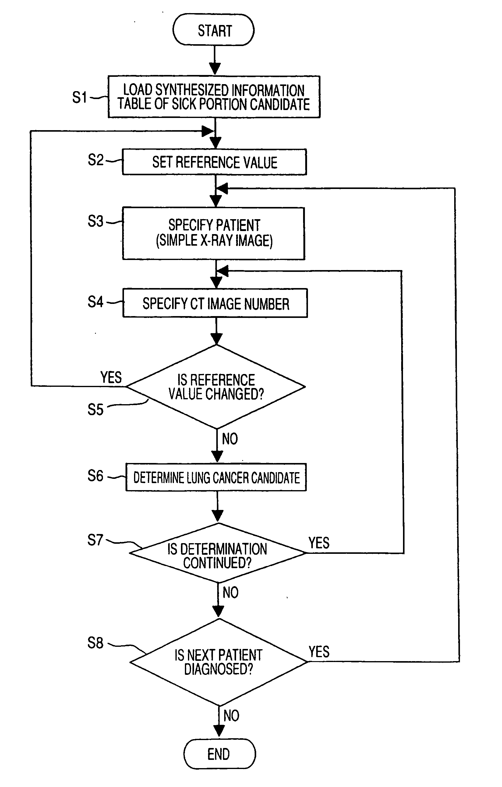
[0046] Referring to the attached drawings, a computer aided diagnostic system according to the invention will be described below.

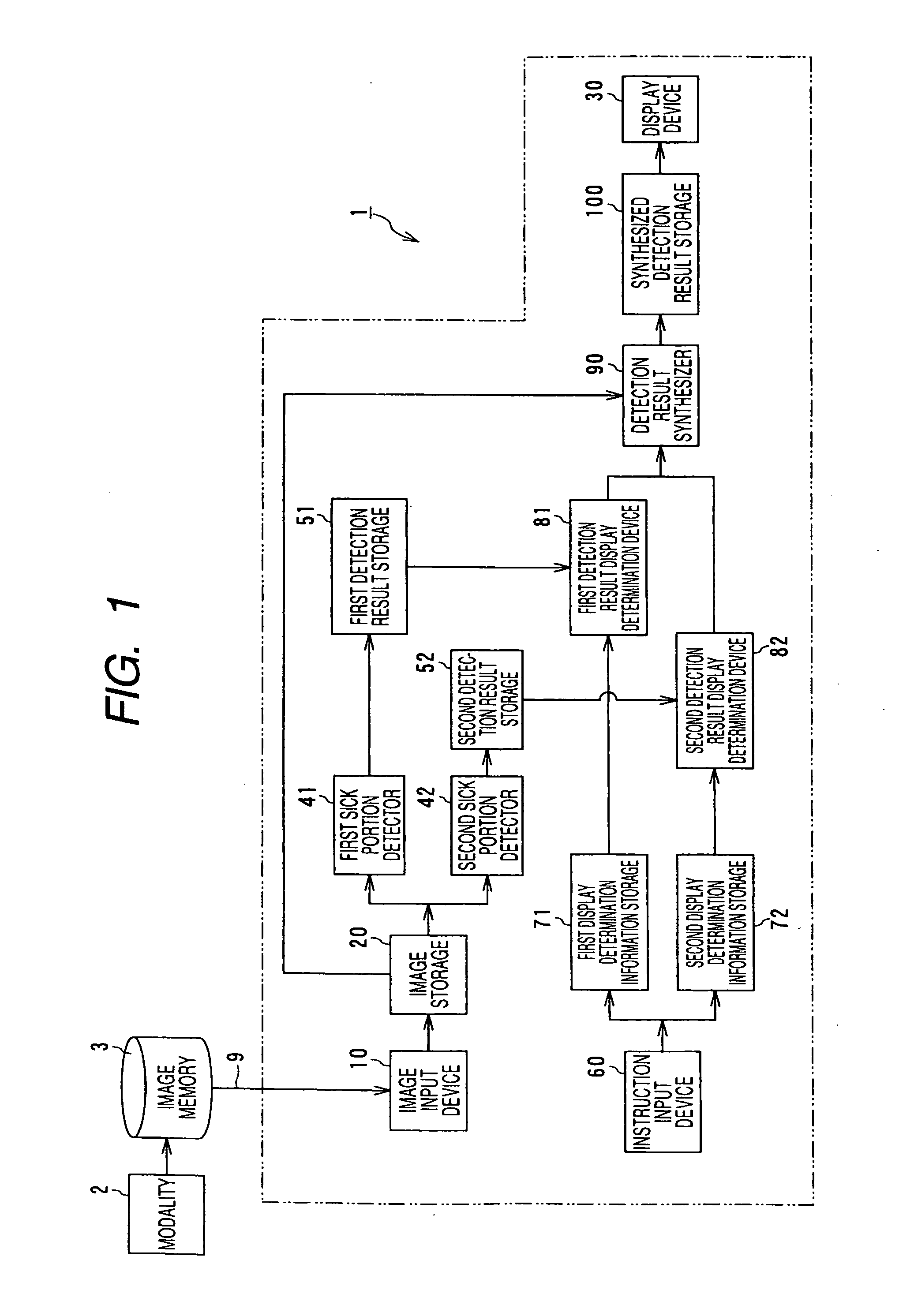
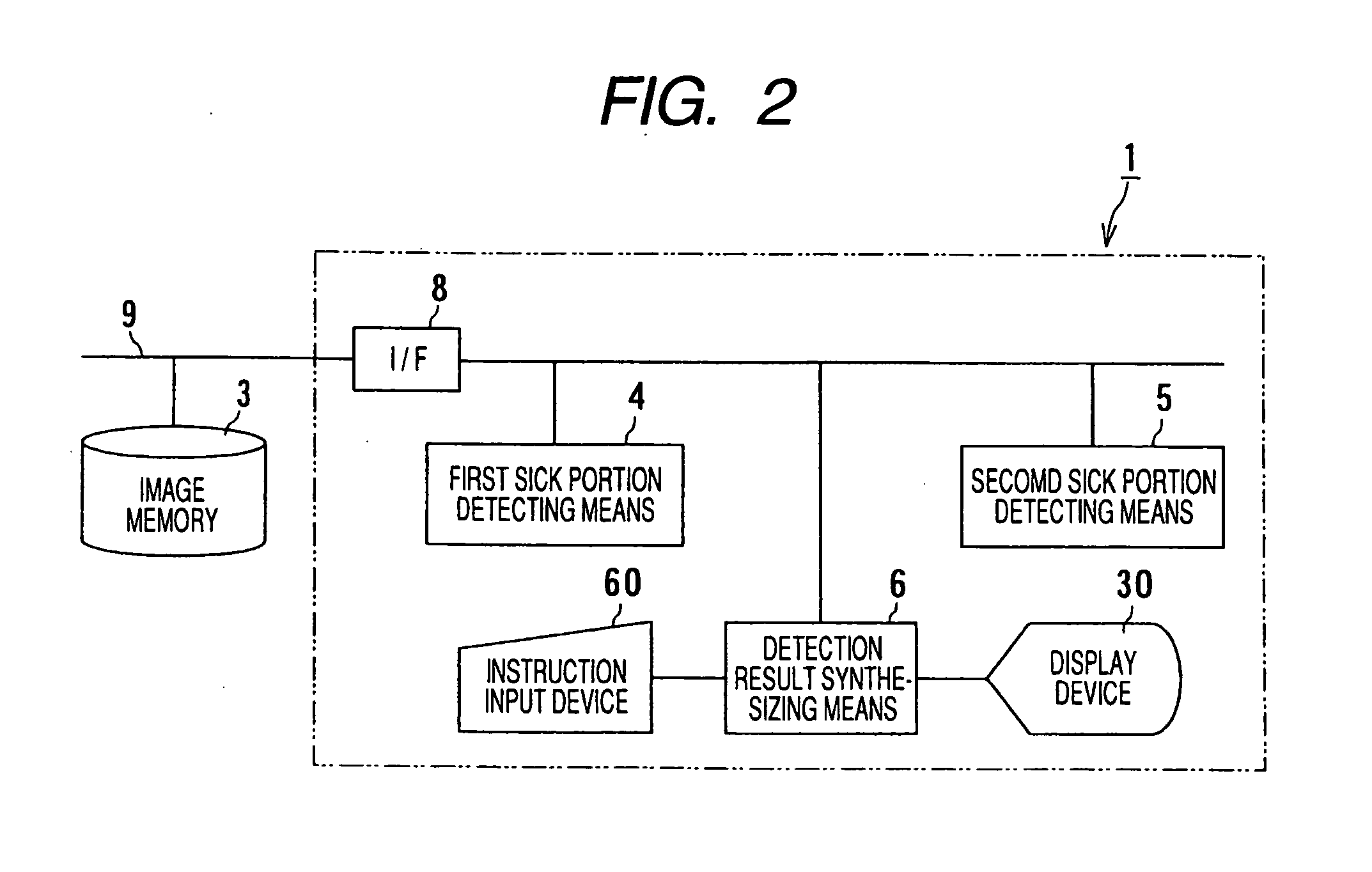
[0047]FIG. 1 is a block diagram showing the outline of a function of the computer aided diagnostic system (a CAD system) 1 equivalent to the first embodiment. An arrow between blocks denotes the flow of main data.

[0048] In the CAD system 1 equivalent to this embodiment, main data input via two input devices, that is, an image input device 10 and an instruction input device 60 is substantially transmitted to first detecting means and second detecting means by grouping the data and after the main data is synthesized by detection result synthesizing means, it is displayed on a display device 30 for displaying the result of detection. In this embodiment, the first detecting means processes and detects an X-ray CT image and the second detecting means processes and detects a simple X-ray image. The above-mentioned simple X-ray image means an X-ray radioscopic i...

second embodiment

[0095] Next, referring to the attached drawings, the CAD system according to the invention will be described.

[0096]FIG. 11 is a block diagram showing the outline of a function of a CAD system 1A equivalent to the second embodiment and FIG. 12 is a block diagram showing an example of the configuration of the CAD system 1A.

[0097] The CAD system 1A equivalent to this embodiment is basically different from that equivalent to the first embodiment in that an image reconstructing device 110 is provided between an image storage 20 and a second sick portion detector 42 as shown in FIG. 11, the other configuration is substantially the same as that in the first embodiment, the same reference numeral is allocated to the same part, and its description is omitted. The image reconstructing device 110 forms image transforming means according to claims. As also clear from FIG. 12, for the configuration of the CAD system 1A, the image transforming means 7 is added to the configuration of the CAD sys...


PUM


Abstract

A computer aided diagnostic system according to the invention is characterized in that first sick portion detecting means for detecting a sick portion candidate based upon an image acquired by a first modality, second sick portion detecting means for detecting a sick portion candidate based upon an image related to the same region of interest of the same subject and acquired by a second modality different from the modality, detection result synthesizing means for comparing the results of detection by the first and second sick portion detecting means and correspondence displaying means for relating the position of the sick portion candidate detected by the first sick portion detecting means on an image analyzed by the second sick portion detecting means and displaying it and for relating the position of the sick portion candidate detected by the second sick portion detecting means on an image analyzed by the first sick portion detecting means and displaying it are provided. According to the above-mentioned configuration, the computer aided diagnostic system that enables a double check comparing images acquired by plural apparatuses can be provided.

Description

TECHNICAL FIELD [0001] The present invention relates to a computer aided diagnostic system for aiding diagnosis using an image for medical application, particularly relates to a computer aided diagnostic system for analyzing plural types of images according to image processing algorithm suitable for respective types by a computer and enabling utilizing them for enhancing the efficiency and reliability of interpreting a shadow by comparing and contrasting acquired abnormality information. BACKGROUND ART [0002] Recently in a medical spot, diagnosis using an imaging diagnostic system (hereinafter called a modality) represented by an X-ray computerized tomograph (X-ray CT), an ultrasonic diagnostic system and a magnetic resonance imaging (MRI) system is frequently performed. Further currently, computer aided detection (CAD) using these is also frequently used (for example, refer to JP-3083606). [0003] The computer aided diagnostic system is a system for automatically extracting sick por...

Claims


Application Information

Patent Timeline
Patent Type & Authority Applications(United States)
IPC IPC(8): G06K9/00G01R33/28A61B5/00A61B5/055A61B6/00A61B6/03G06T1/00
CPCA61B6/032A61B6/5235A61B6/465A61B6/463A61B5/0035A61B5/7425G06T7/0012G06T2207/30064
Inventor OZAKI, MASAHIROIWATA, ITSUKO
Owner TOSHIBA MEDICAL SYST CORP
Why Eureka
Try Eureka