System and method for tracking anatomical structures in three dimensional images

a three-dimensional image and anatomical structure technology, applied in image enhancement, instruments, applications, etc., can solve the problems of inability to recover accurate regional motion, inability to measure data, and inability to accurately detect the movement of the body

Inactive Publication Date: 2006-03-23
SIEMENS HEALTHCARE GMBH
View PDF7 Cites 100 Cited by
  • Summary
  • Abstract
  • Description
  • Claims
  • Application Information

AI Technical Summary

Problems solved by technology

For most visual tracking applications, measurement data are uncertain and sometimes missing: images are taken with noise and distortion, while occlusions can render part of the object-of-interest unobservable.
Ultrasound is prone to reflection artifacts, e.g., specular reflectors, such as those that come from membranes.
However, most existing segmentation or detection methods do not attempt to recover accurate regional motions of the endocardial wall, and in most cases, motion components along the wall are ignored.
This is not suitable for regional wall abnormality detection, because regional motion of an abnormal left ventricle is likely to be off the normal of the contour, not to mention that global motion, such as translation or rotation (due to the sonographer's hand motion or respiratory motion the patient), causes off-normal local motion on the contour as well.

Method used


Image

  • System and method for tracking anatomical structures in three dimensional images
  • System and method for tracking anatomical structures in three dimensional images
  • System and method for tracking anatomical structures in three dimensional images

Examples


Embodiment Construction

[0022] The present invention is directed to a system and method for tracking three dimensional motion of an anatomical structure. An example where such a method would be utilized is for detecting regional wall motion abnormalities in the heart by diction and segmentation of the ventricle endocardial or epicardial borders through machine learning, or classification, and by identifying similar cases from annotated databases. It is to be understood by those skilled in the art that the present invention may be used in other applications where motion tracking is useful such as, but not limited to, surveillance. The present invention can also be used in 4 dimensional (3D+time) data analysis, such as medical analysis of anatomical structures such as the heart, lungs or tumors, which can be evolving over time.

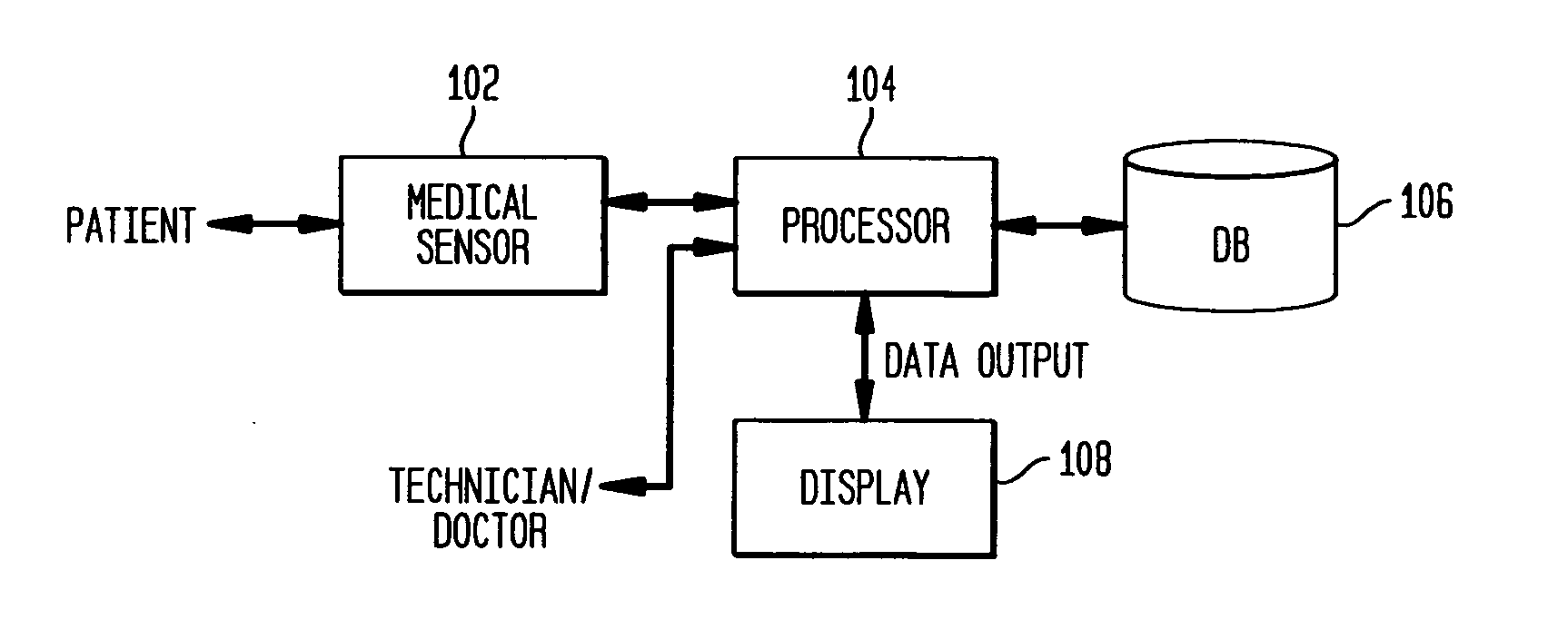
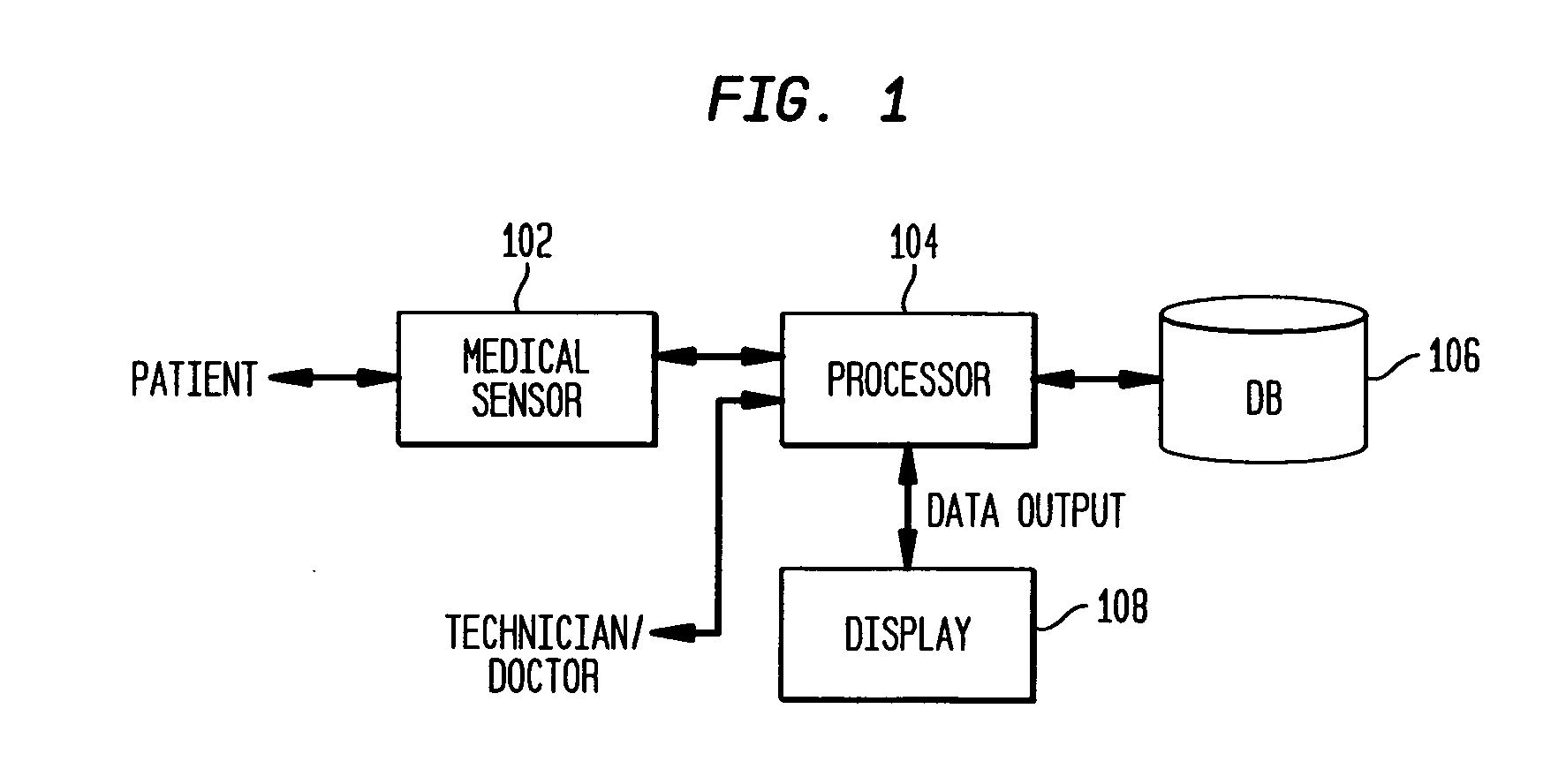
[0023] For purposes of describing the present invention, an example will be described for detecting the endocardial wall of the left ventricle of a human heart. FIG. 1 illustrates an ...


PUM


Abstract

A system and method for defining and tracking a deformable shape of a candidate anatomical structure wall in a three dimensional (3D) image is disclosed. The shape of the candidate anatomical structure is represented by a plurality of labeled 3D landmark points. At least one 3D landmark point of the deformable shape in an image frame is defined. A 3D cuboid is defined around the detected 3D landmark point. For each landmark point associated with the anatomical structure, its location and location uncertainty matrix is estimated in subsequent frames relative to the reference anatomical structures. A shape model is generated to represent dynamics of the deformable shape in subsequent image frames. The shape model includes statistical information from a training data set of 3D images of representative anatomical structures. The shape model is aligned to the deformable shape of the candidate anatomical structure. The shape model is fused with the deformable shape. A current shape of the candidate anatomical structure is estimated.

Description

CROSS-REFERENCE TO RELATED APPLICATION [0001] This application claims the benefit if U.S. Provisional Application Ser. No. 60 / 606,635, filed Sep. 2, 2004, which is incorporated by reference in its entirety.FIELD OF THE INVENTION [0002] The present invention is directed to a system and method for tracking anatomical structures in three dimensional images, and more particularly, to a system and method for tracking motion of anatomical structures in three dimensional images by monitoring trajectories of landmark points associated with the anatomical structure. BACKGROUND OF THE INVENTION [0003] It is very common during medical examinations for medical imaging systems (e.g., ultrasound imaging systems) to be used for the detection and diagnosis of abnormalities associated with anatomical structures (e.g., organs such as the heart). Many times, the images are evaluated by a medical expert (e.g., a physician or medical technician) who is trained to recognize characteristics in the images ...

Claims


Application Information

Patent Timeline
Patent Type & Authority Applications(United States)
IPC IPC(8): A61B5/05
CPCA61B8/08A61B8/0883A61B8/463A61B8/483G06K9/3216G06K2209/051G06T2207/30048G06T7/0089G06T7/2033G06T7/208G06T2200/04G06T2200/24G06T2207/10136G06T7/0083G06T7/0016Y10S128/922G06T7/12G06T7/149G06T7/246G06T7/277G06V10/245G06V2201/031
Inventor COMANICIU, DORINGEORGESCU, BOGDANXIAO, JINGZHOU, XIANG
Owner SIEMENS HEALTHCARE GMBH
Why Eureka
Try Eureka