Method for the separation of cell fractions

Inactive Publication Date: 2006-08-03
LEHNERT LASSE
View PDF1 Cites 10 Cited by
  • Summary
  • Abstract
  • Description
  • Claims
  • Application Information

AI Technical Summary

Benefits of technology

[0030] 3) It must be possible to perform the method with a minimum of effort at the place of sampling in a reliable manner so that the method is standardized. Standardized in this context means firstly that the systems used for sampling must lend themselves to pre-fabrication. On the other hand, it is necessary for the sampling protocol to be devised such that the method can be performed by different investigators with equal efficiency when proceeding in the same manner. By definition, it is not possible to use a te

Problems solved by technology

It was found that incubation in a hypotonic solution results in

Method used


Image

  • Method for the separation of cell fractions
  • Method for the separation of cell fractions
  • Method for the separation of cell fractions

Examples


example 1

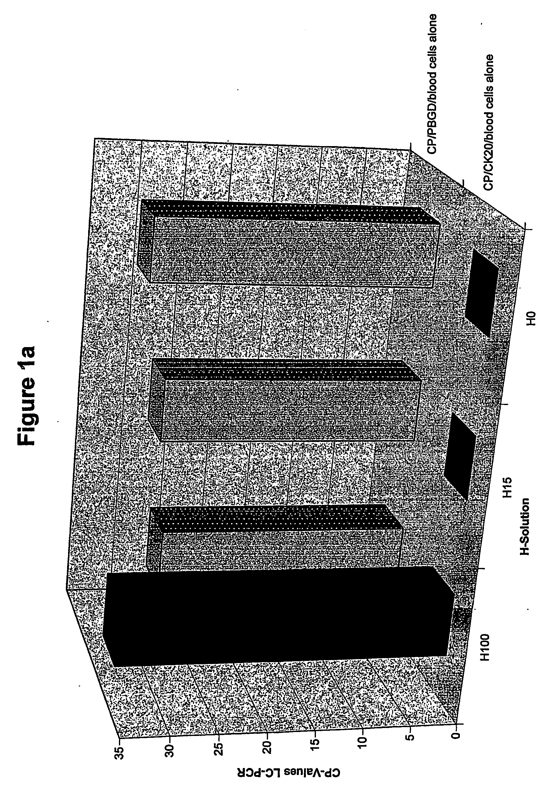
[0116] Blood was taken from a blood donor and divided into 2 aliquots. The first aliquot was treated on its own and for the second aliquot after addition of 1000 HT29 tumor cells. Since only 20% of the cDNA generated can be used for the CK-20 PCR, the signals correspond to those for 200 HT29 cells. The samples were treated and analyzed in the same manner as the reference example.

[0117]FIG. 1a shows the results for the test run without addition of tumor cells. CK20 and PBGD signals are detected for the blood cells treated with a solution of 276 mosmoles (H100). No CK20 signal is seen after treatment with H solutions (H15-HO) with a lower osmolality (<46 mosm / kg), but the PBGD signal is present (sufficient sample material was therefore present).

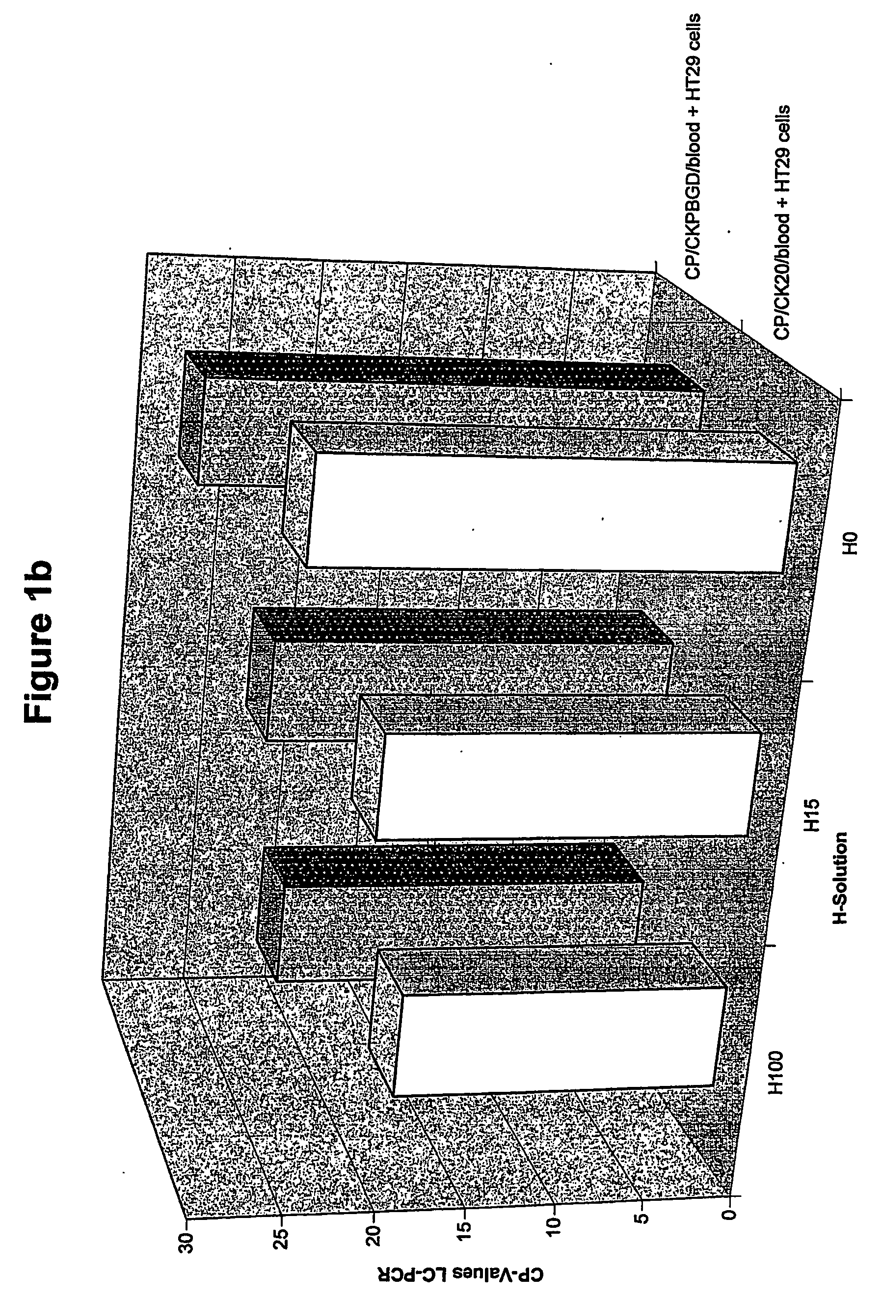
[0118]FIG. 1b shows the results for the test run with tumor cells added (HT29). CK20 and PBGD signals are also detected after treatment of the sample with solutions of low and very low osmolality (H15, HO). Since no CK20 signal was seen in th...

example 2

[0125] Blood was taken from a blood donor and split into 2 aliquots. The first aliquot was used without addition of tumor cells the second aliquot was mixed with 25 HT29 tumor cells.

[0126] The samples were prepared and analyzed in the same manner as the reference example.

[0127] Since only 20% of the cDNA generated can be used for the CK-20 PCR, the s signals correspond to those for 5 HT29 cells. For this donor, the background signal was reached with hypotonic solutions at an osmolality of just 103 mosm / kg.

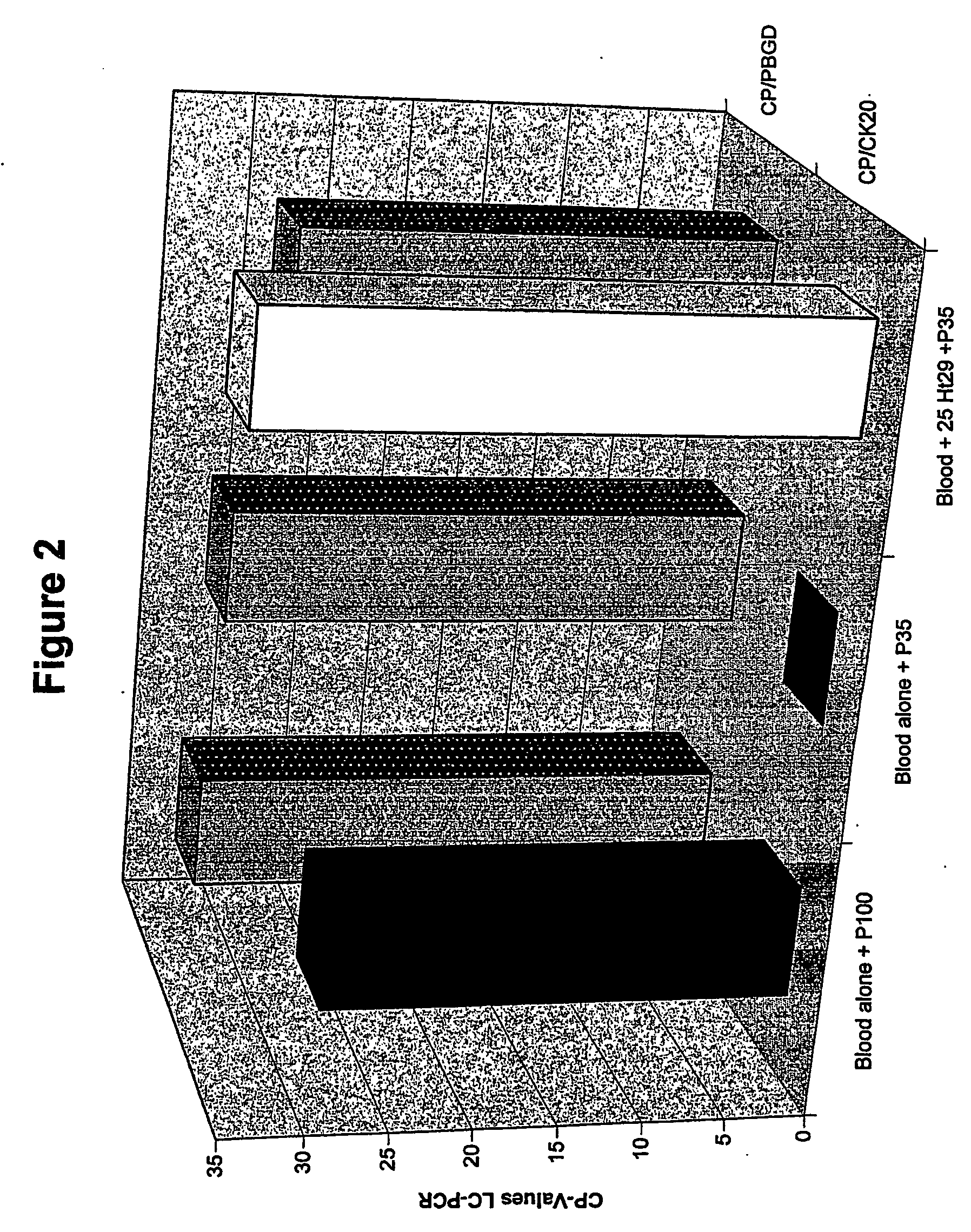
[0128]FIG. 2 shows the results of quantitative RT-PCR for CK20 and PBGD. The CK20 and PBGD signals can be detected without treatment of the blood cells with hypotonic solutions (blood alone). Incubation with a P35 solution (103 mosm / kg) leads to elimination of the CK20 signal. The PBGD signal is retained (with P35). In the parallel run, containing 25 HT29 tumor cells, the CK20 signal remains detectable (blood+25Ht29+P35).

example 3

Improvement of the Results of Lysis Through Addition of RNase A

[0129] Example 1 was repeated. RNase A (1 mg / aliquot) was added to the blood before treatment with the P / H solution to enable rapid degradation of the RNA before the final lysis through the RLT buffer containing guanidinium isothiocyanate. This led to an improvement in the results of lysis, i.e., lysis was also possible with solutions of higher osmolality.


PUM

PropertyMeasurementUnit
Weightaaaaaaaaaa
Weightaaaaaaaaaa
Weightaaaaaaaaaa

Abstract

The present invention relates to a method for the separation of cells, in particular for the preparation of samples in tumor diagnostics. In particular, the present invention relates to a method for sample preparation for the detection of tumor cells of solid tumors in the course of the diagnosis for prognosis and stratification of therapy, comprising the destruction of cells that make this diagnosis more difficult or entirely impossible. Furthermore, the present invention relates to a kit for the preparation of samples and for the detection of the presence of the altered cells. Finally, the present invention relates to the use of the method and the kit in the diagnostics of altered cells such as tumor cells, and the performance of a PCR reaction to detect the tumor cells in body fluids and tissues.

Description

[0001] The present invention relates to a method for the separation of cells, in particular for the preparation of samples in tumor diagnostics. More exactly, the present invention relates to a method for sample preparation for the detection of tumor cells of solid tumors in the course of diagnostics for prognosis and stratification of therapy, embracing the destruction of cells that make this diagnosis more difficult or entirely impossible. Furthermore, the present invention relates to a kit for the preparation of samples and for the detection of the presence of the altered cells. Finally, the present invention relates to the use of the method and the kit in the diagnostics of altered cells such as tumor cells, and the performance of a PCR reaction to detect the tumor cells in body fluids and tissues. PRIOR ART [0002] The separation of different cell populations is a central aspect in the analysis of different cell populations. Very different methods are known in the prior art for ...

Claims


Application Information

Patent Timeline
IPC IPC(8): C12Q1/68C12N5/08C12N5/02C12N5/06
CPCC12Q1/6886C12Q2600/158
Inventor LEHNERT, LASSE
Owner LEHNERT LASSE
Why Eureka
Try Eureka