
Miniature ingestible capsule

a capsule and small technology, applied in the field of small ingestible capsules, can solve the problems of low treatment cost, health care population at risk of invasive procedure, and inability to perform colorectal cancer screening for the vast majority of the population, so as to reduce the need for highly trained personnel, economic affordability, and less expensive

Active Publication Date: 2006-08-31
NAIR PADMANABHAN P +1
View PDF33 Cites 146 Cited by
  • Summary
  • Abstract
  • Description
  • Claims
  • Application Information

AI Technical Summary

Benefits of technology

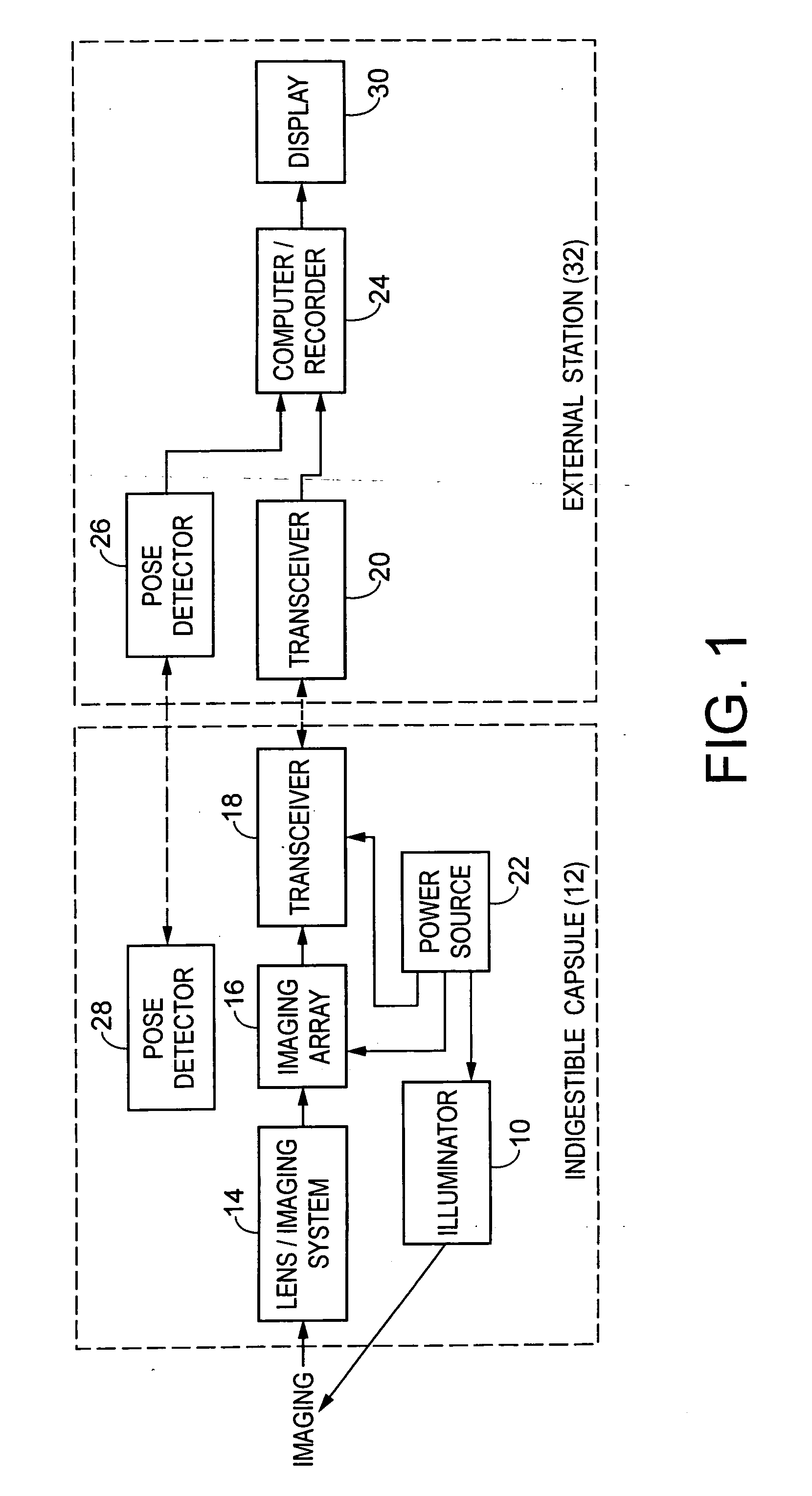
[0022] In another form, the capsule includes a reception capability, such as a radio-frequency receiver, and an internal microprocessor that allows instructions to be relayed from the physician to the capsule. Miniature motors allow the imaging system to be reoriented, or provide some form of “controlled mobility,” which could include a remote control, joystick, mouse control or other computer-directed or voice-directed control or other control. An expandable bladder attached to the capsule can be expanded to stabilize the capsule or slow its motion through the tract. The system may also include on-board signal processing circuitry to automatically stabilize the image. Alternatively, a micro-machined mechanical stabilization platform can be built into the imaging system. The imaging system may also include a means such as a prism or fiber-optic device, to direct multiple images onto the imaging device.
[0028] Another aspect of the present invention is that it would eliminate the need for highly trained personnel for its operation.
[0029] Yet another aspect of the present invention is that it is economically affordable and less expensive than existing methods of imaging gastrointestinal tracts.
[0030] Still another aspect of the present invention is that it is easier to implement and allows patients to be examined more frequently than existing methods of imaging gastrointestinal tracks.
[0031] Still other functions and benefits of an embodiment of the invention include the ability to provide ultrasound imaging with a miniature ultrasound sensor and to image and treat problems with the pancreatic and biliary ducts and gastrointestinal system and the rest of the human body. In another embodiment, with a special dye injection port for various dyes imaging of, for example, the biliary and pancreatic ducts and the rest of the human body can be done. Treatment apparatus ports allow for cutting and coagulation currents to be delivered locally.
[0032] Furthermore, a focal laser treatment, argon plasma coagulator treatment, lithotripsy treatment with ultrasound current can be used in the biliary and pancreatic ducts, gastrointestinal system and rest of human body in other forms of the invention. In addition, tools including miniature baskets, miniature snares, miniature needles with epinephrine (for example) and other treatments, miniature forceps, miniature cytology brushes can be included in a form of the capsule for diagnosis and treatment in the biliary and pancreatic trees, pancreas, liver, and gastrointestinal system, and human body. Also, a thin wire, catheter, miniature plastic or metal stint can be deployed in the biliary and pancreatic ducts in other forms of the capsule. Finally, the capsule has suction capabilities to remove unwanted debris and can sprinkle water and n-acetyl cysteine locally over the lens or capsule to remove residual debris from the gastrointestinal system to improve or enhance visualization, diagnostic, therapeutic or other functions of the capsule.

Problems solved by technology

Survival rates improve and treatment costs decline with early detection of the process.
However, regular screening for colorectal cancer is not performed for the vast majority of the populace due to the high cost of such programs and, more importantly, the reluctance of a healthy population at risk to undergo an invasive procedure again and again for surveillance against cancer.
These tests exhibit poor sensitivity due to the fact that malignant growths of the colon have to be fairly large before they start to bleed.
Furthermore, there are many other reasons for bleeding into the gastrointestinal tract (e.g., ulcers) which lead to low specificity of the test and a high probability of false positives.
Considering the cost of the colonoscopy alone, a yearly colonoscopy for all patients over age 48 for instance, would be prohibitively expensive.
Although simpler than a colonoscope, its operation still requires the presence of a highly trained physician and often requires sedation.
Again, its operation requires the presence of a highly trained physician and often requires sedation.
Considering the cost of endoscopy and the sedation requirement, it would be prohibitively expensive to perform esophagogastroduodenoscopy on all patients with symptoms.
Their operation requires the presence of a highly trained physician and a lot of sedation.
More sophisticated approaches such as colonoscopy and related gastrointestinal imaging methods, namely, sigmoidoscopy and esophagogastroduodenoscopy, are more effective because they can identify abnormalities before the occurrence of late-stage symptoms (e.g., blood in the stools for colonic tumors or tarry stools for peptic ulcers) However, these methods see limited use for several reasons.
One, they are invasive and uncomfortable to the patient, requiring sedation so that a flexible fiberoptic tube can be inserted into the tract.
This is a major limitation of these tests in their application to healthy asymptomatic individuals for repeated examinations (every 1-3 years).
Secondly, these tests are expensive, requiring the presence of a physician and other personnel.
Third, they are inconvenient, requiring the patient to take a purgative, fast overnight, and remain incapacitated during the procedure.

Method used


Image

  • Miniature ingestible capsule
  • Miniature ingestible capsule
  • Miniature ingestible capsule

Examples


fourth embodiment

[0071] Referring now to FIG. 6, wheels 140 may be added to the capsule 150 in accordance with the present invention. The capsule may be one of the three embodiments previously described. The wheels would be powered by a remote control device (not shown). The wheels 140 would have spokes 142 which would provide friction and enable the wheels to move along surfaces. The capsule with wheels would be used primarily for situations where the stomach is virtually dry, not fluid filled, such as a result of the patient fasting. Two sets of four wheels 140 equally spaced may be placed around the circumference of the capsule. Also, the capsule with wheels may be used in addition to a second capsule. The capsule with wheels may provide a therapeutic function instead of a diagnostic function. That is, the capsule may supply medicine or perform another therapeutic function to the area being investigated by the second capsule. One capsule would not be able to detect the area being investigated thr...

fifth embodiment

[0072] In the situation where the stomach is at least partially fluid-filled, fins 160 in lieu of wheels may be placed on the exterior surface of capsule 170 as illustrated in a fifth embodiment in FIG. 7. The fins 160 would aid the capsule in moving throughout the stomach.

sixth embodiment

[0073] In a sixth embodiment, referring to FIG. 8, prongs 180 would extend from posterior end 182 of capsule 184. The prongs 180 can be retractable and would serve as a base for the capsule to effectively anchor or stabilize the capsule. A laser or biopsy forceps (not shown) could extend from the anterior portion 186 of the capsule.


PUM


Abstract

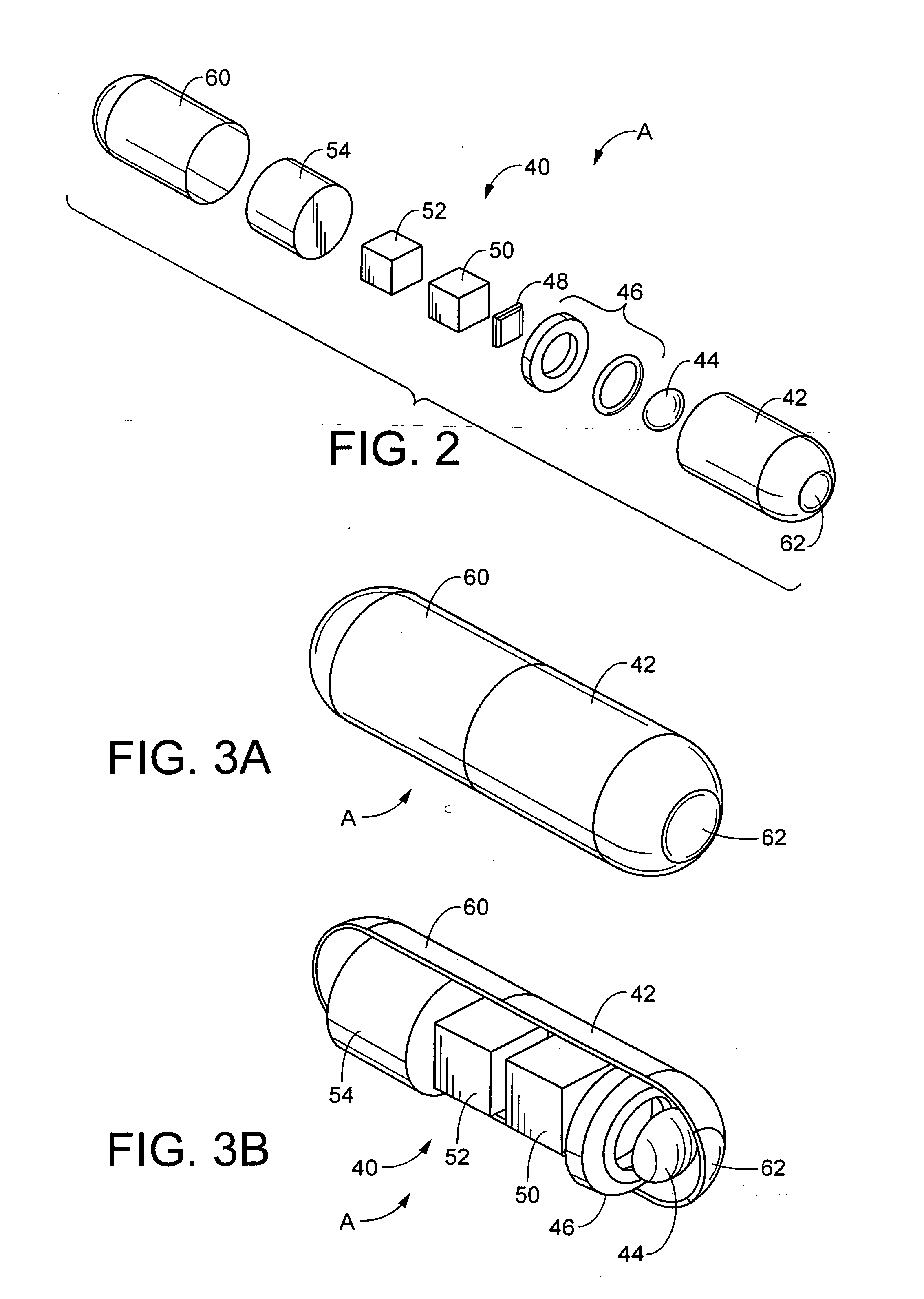
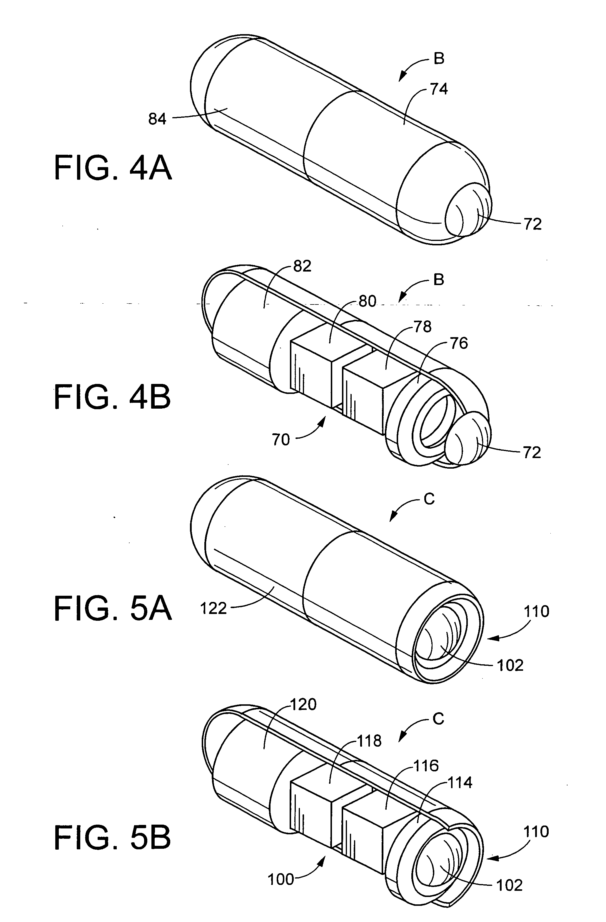
A miniature ingestible capsule has multiple therapeutic or diagnostic operations that can be performed. These functions are controlled by a combination of an outside control, a pose beacon and through information relayed from an imagining array and transmitter. These functions can be in a separate capsule without an imaging array or within the same capsule with an imaging array. Typically, there is one function performed in addition to imaging. These functions can include suction and spray capabilities, ultrasound sensor, lithotripsy, laser, heat, electrocautery, BICAP, biopsy forceps, a needle knife snare cautery (cold and hot with continuous or pulsed current for cutting and coagulation), with a basket, and fine needle aspiration with various wheels and fins and motors controlled externally and other tools to be used in humans. All of these tools can be attached to a retractable arm. Also, they can be used on an elevator device that lifts them, allowing for an extra 180° of movement.

Description

[0001] This application claims priority from U.S. Provisional Application Ser. No. 60 / 180,960, filed on Feb. 8, 2000.FIELD OF THE INVENTION [0002] This invention relates to a miniature ingestible capsule for imaging the gastrointestinal tract for medical diagnosis or diagnosis and / or therapy for the human body. More specifically, this invention relates to noninvasive, noninterventional methods for internal examination of the gastrointestinal tract or other internal therapy and diagnosis of the human body that are significantly more convenient, comfortable, lower in cost and more advanced compared with current invasive diagnostic methods such as colonoscopy, sigmoidoscopy, esophagogastroduodenoscopy and push enteroscopy. BACKGROUND OF THE INVENTION [0003] The mammalian gastrointestinal tract comprises the esophagus, stomach, small intestine, and colon. Physicians image the interior of the gastrointestinal tract to aid in the diagnosis and treatment of many illnesses such as ulcers, g...

Claims


Application Information

Patent Timeline
Patent Type & Authority Applications(United States)
IPC IPC(8): A61B1/00A61B1/05A61B1/12A61B5/07A61B8/12
CPCA61B1/00016A61B1/00082A61B1/00087A61B1/00094A61B1/00156A61B1/041A61B1/12A61B1/126A61B5/06A61B5/061A61B5/073A61B5/411A61B5/4839A61B5/6882A61B8/12
Inventor MULLICK, TARUNNAIR, RAMGOPALDUTTA, SUDHIR K.NAIR, PADMANABHAN P.
Owner NAIR PADMANABHAN P
Learn More