Method for movement-compensation in imaging

a technology of movement compensation and imaging, applied in the field of movement compensation in imaging, can solve the problems and reducing the number of events measured per individual image. , to achieve the effect of reducing the number of events measured per individual image and reducing the diagnostic value of images

Active Publication Date: 2006-10-19
SIEMENS HEALTHCARE GMBH
View PDF6 Cites 31 Cited by
  • Summary
  • Abstract
  • Description
  • Claims
  • Application Information

AI Technical Summary

Benefits of technology

[0008] The disadvantage of the method that involves ECG gating is that the number of events measured per individual image is reduced as a result of the di...

Problems solved by technology

The disadvantage of the method that involves ECG gating is that the number of events measured per individual image is reduced as a result of the division into time intervals.
This has a detrimental eff...

Method used


Image

  • Method for movement-compensation in imaging
  • Method for movement-compensation in imaging

Examples


Embodiment Construction

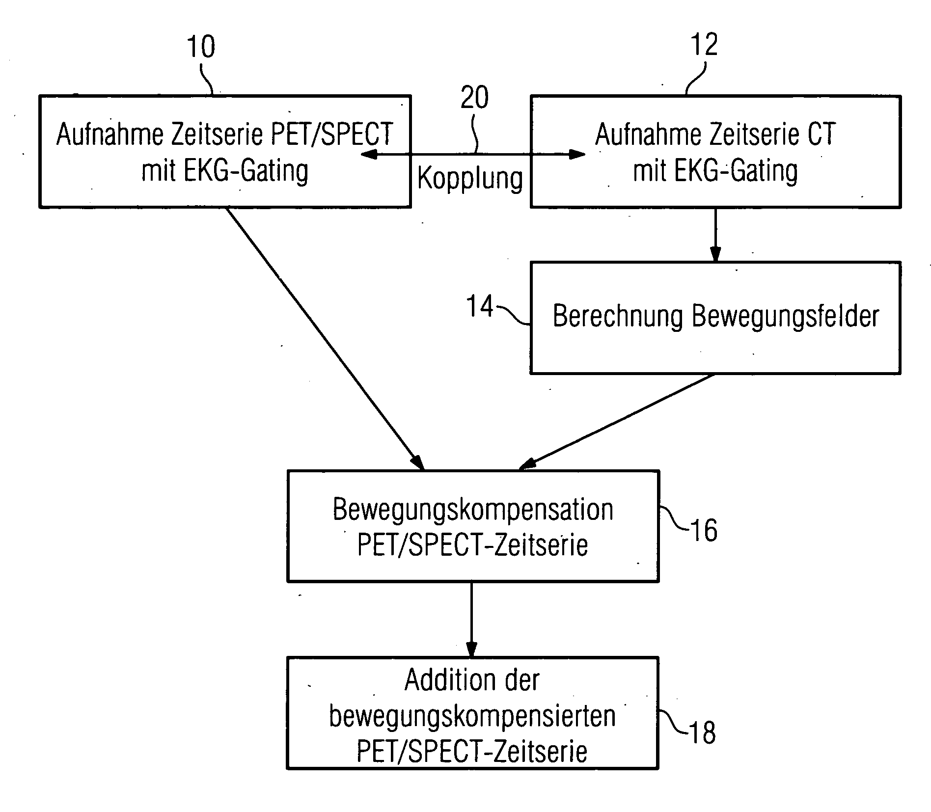
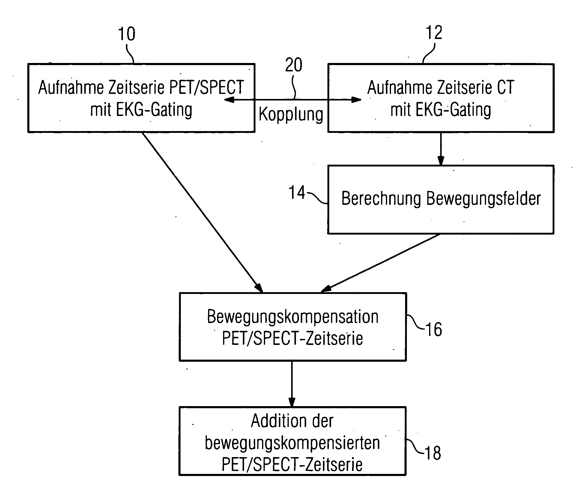
[0024] In the preferred embodiment of the invention, a person's heart is being investigated. A combined PET-CT-System or SPECT-CT-system is used, wherein both the parts of the system are mechanically connected to each other, su ch that the images that are generated with the PET part of the system can be directly correlated with the CT images.

[0025] Furthermore, the option of carrying out ECG gating is available, that is, ECG probes can be attached to the patient, and an image evaluation analyses the ECG signal that has been recorded. The ECG signal is divided into a plurality of intervals, each interval corresponding to a specific image from a time series. The system now records image data and, at the same time, the ECG curve. On the basis of the interval in which the heart is located during the recording of the image data, the image data are assigned in the respective interval and in the respective partial image from the time series of image s. Finally a time series of three-dimen...


PUM


Abstract

In a method for mathematical compensation of a periodic movement of an organ in a first image-generation method used to image said organ, two time series of three-dimensional image data are acquired using gating, one by the first image-generation method and one by the second image-generation method, the image data that have been acquired by the second image-generation method being used to calculate motion fields which are applied for the compensation of the data from the time series which was acquired by the first image-generation method. The compensation encompasses the mathematical inclusion of motion fields and the mapping of the image data to a reference time. All the image data mapped back to the reference time are added together.

Description

CROSS REFERENCE TO RELATED APPLICATIONS [0001] This application claims priority to the German Application No. 10 2005 017 492.2, filed Apr. 15, 2005 which is incorporated by reference herein in its entirety. FIELD OF INVENTION [0002] The invention relates to a method for mathematical compensation of a periodic movement of an organ in a first image generation method used for imaging said organ. [0003] It also relates to an image recording system for a first image generation method. BACKGROUND OF INVENTION [0004] If images are taken of the heart by means of nuclear medical investigation methods such as, for example, PET (Positron Emission Tomography) and SPECT (Single Positron Emission Computer Tomography), this involves long acquisition times typically lasting a few minutes (as in PET) and up to more than 20 minutes (in SPECT). The resulting data set therefore represents a time-related averaging over all the cardiac phases and the cardiac motion therefore appears blurred. [0005] Furt...

Claims


Application Information

Patent Timeline
IPC IPC(8): A61B5/05
CPCA61B5/7285A61B6/5264A61B6/527G06T7/20A61B8/5276A61B8/543A61B6/541
Inventor BOESE, JANKLEEN, MARTINPFISTER, MARCUSRAHN, NORBERT
Owner SIEMENS HEALTHCARE GMBH
Why Eureka
Try Eureka