Automatic determination of the standard cardiac views from volumetric data acquisitions

a volumetric data and automatic determination technology, applied in the field of automatic determination of standard cardiac image views from volumetric data, can solve the problems of computational cost and computational cost of em algorithms, and achieve the effect of accurate estimation of standard cardiac image views

Inactive Publication Date: 2006-10-26
SIEMENS MEDICAL SOLUTIONS USA INC
View PDF34 Cites 31 Cited by
  • Summary
  • Abstract
  • Description
  • Claims
  • Application Information

AI Technical Summary

Benefits of technology

[0010] The present invention provides methods and a system to accurately estimate standard cardiac image views as defined by the American Heart Association from a 3D volume provide by one of several possible imaging techniques. It is one aspect of the present invention to provide methods for automatic segmentation of the Left Ventricle in a 3D image of the heart and based on the LV segmentation to estimate the SA, LHA and VLA for standard cardiac image views.

Problems solved by technology

This is computationally expensive and assumes a fixed field of view.
EM algorithms are also computationally expensive.

Method used


Image

  • Automatic determination of the standard cardiac views from volumetric data acquisitions
  • Automatic determination of the standard cardiac views from volumetric data acquisitions
  • Automatic determination of the standard cardiac views from volumetric data acquisitions

Examples


Embodiment Construction

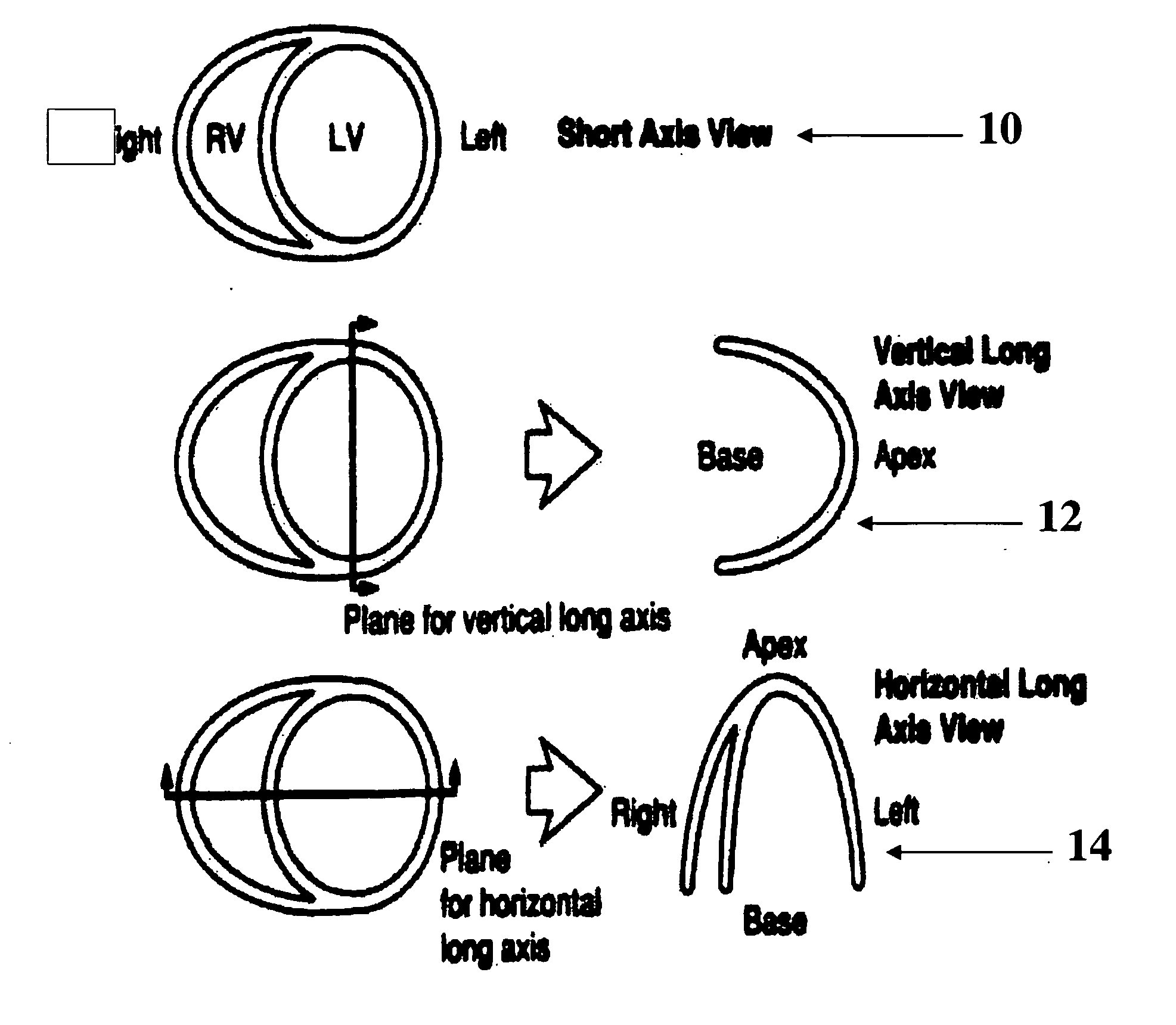
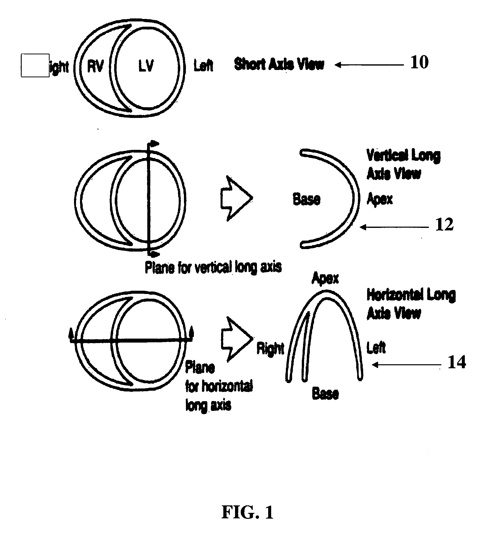
[0032] In the following, volumetric data, as obtained from known medical-imaging equipment, is worked upon to present to a user a visually useful image corresponding to the volumetric data. The volumetric data may be obtained, for example, from whole heart magnetic resonance angiography (MRA). It will be appreciated that volumetric data obtained by other means may also be utilized. That is, the present invention is not limited to specific types of volumetric data, file formats, voxel or pixel resolutions, or the like. The volumetric data may be thought of as describing a plurality of specific locations in space, each with a corresponding intensity value. Of course, the volumetric data may contain additional information, but such additional information is not required for the purposes of the following disclosure.

[0033] The present invention method may be implemented in the form of a computer program executable on any suitable computing device, such as a personal computer, as known i...


PUM


Abstract

A system and method for automatically determining the standard cardiac image views as defined by the American Heart Association from volumetric data of the chest including the heart. The system and method can be used by a health practitioner to quickly see the two dimensional views from which a diagnosis is generally made. The left ventricle is detected. Then the relative orientation of the right ventricle is determined and the standard cardiac views are determined.

Description

CROSS-REFERENCE TO RELATED APPLICATIONS [0001] This application claims the benefit of U.S. Provisional Application No. 60 / 665,160, filed Mar. 25, 2005, which is incorporated herein by reference.BACKGROUND OF THE INVENTION [0002] The present invention relates to processing systems and methods for automatically determining predefined viewing directions of a heart from medical imaging data. [0003] The standard views for the display of cardiac-related imagery are defined by the American Heart Association. These views include the short axis view, the vertical long axis view, and the horizontal long axis view. With reference to FIG. 1, the standard views may be defined in relation to the left ventricle 12 and right ventricle 14 of a heart 10, and include a short axis view (SA), a vertical long axis view (VLA) and a horizontal long axis view (HLA). For orientation reference FIG. 2 provides a schematic view of the heart and FIG. 3 provides a view of an image slice with right and left ventri...

Claims


Application Information

Patent Timeline
Patent Type & Authority Applications(United States)
IPC IPC(8): G06K9/34
CPCG06T7/0081G06T19/00G06T2207/10088G06T2210/41G06T2207/20156G06T2207/30048G06T2207/20092G06T7/11
Inventor SUN, YINGGRADY, LEOFUNKA-LEA, GARETHO'DONNELL, THOMAS
Owner SIEMENS MEDICAL SOLUTIONS USA INC
Why Eureka
Try Eureka