Multiple transducer configurations for medical ultrasound imaging

a multi-configuration, ultrasound imaging technology, applied in the field of medical ultrasound imaging systems, can solve the problem of complex image quality produced at different depths

Inactive Publication Date: 2006-11-09
SCI MED LIFE SYST
View PDF42 Cites 143 Cited by
  • Summary
  • Abstract
  • Description
  • Claims
  • Application Information

AI Technical Summary

Problems solved by technology

However, the level of image quality produced at different depths is a complex interplay of numerous factors, such as overall system bandwidth (for example, the bandwidth of the receiving circuitry), transducer focus, beam pattern in addition to transducer frequency.

Method used


Image

  • Multiple transducer configurations for medical ultrasound imaging
  • Multiple transducer configurations for medical ultrasound imaging
  • Multiple transducer configurations for medical ultrasound imaging

Examples


Embodiment Construction

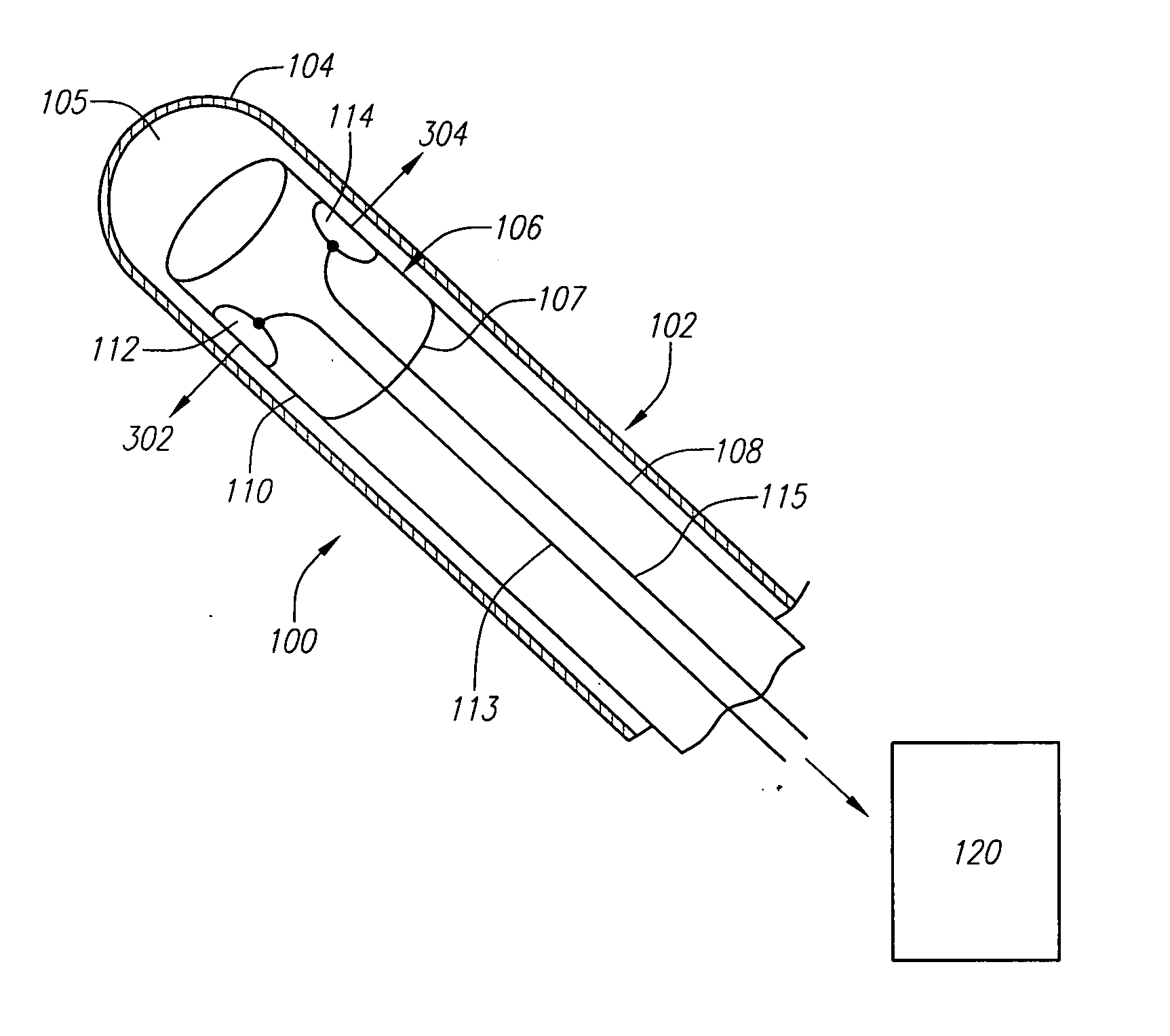
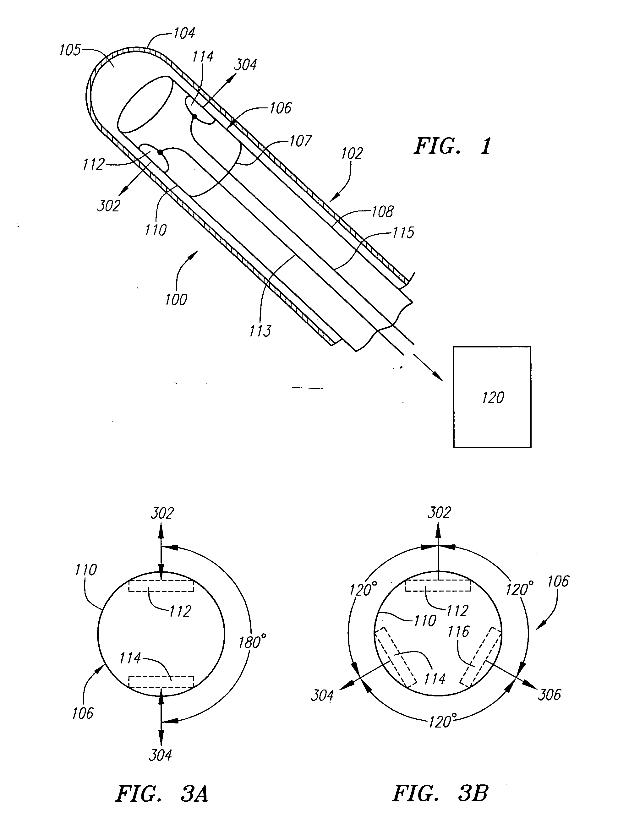
[0025] The systems and methods described herein provide for multiple transducer configurations in ultrasound imaging systems. These systems and methods allow an ultrasound imaging system to image a greater range of tissue depths while maintaining a relatively high degree of image quality. FIG. 1 depicts a schematic diagram of one example embodiment of an ultrasound imaging system 100 for use with the systems and methods described herein. Preferably, imaging system 100 is an IVUS imaging system, although the systems and methods are not limited to such and any other type of imaging system, such as ICE, can be used. Here, catheter 102 is shown having elongate tubular outer sheath 104 and inner lumen 105. An imaging device 106 is preferably mounted on distal end 107 of rotatable driveshaft 108, which is configured to move, or slide, within inner lumen 105. System 100 is preferably configured to image a tissue cross-section by rotating imaging device 106, although system 100 is not limit...


PUM


Abstract

The systems and methods described herein provide for multiple transducer configurations for use in medical ultrasound imaging systems. A medical device has a rotatable imaging device located therein for imaging an internal body lumen or cavity. The imaging device can include multiple transducers each configured to image a separate tissue depth or range of tissue depths. The transducers can be configured to operate over separate frequency ranges, with separate physical focuses or any combination thereof. Also provided is an image processing system configured to combine the image data collected from each transducer into a tissue image.

Description

FIELD OF THE INVENTION [0001] The systems and methods relate generally to medical ultrasound imaging systems and, more particularly, to multiple transducer configurations for imaging wider depth ranges. BACKGROUND INFORMATION [0002] Conventional medical ultrasound imaging systems, such as intravascular ultrasound (IVUS) and intracardiac echocardiography (ICE), use an ultrasound imaging device to image the interior of a living being. The ultrasound imaging device is placed on or within a catheter, which can then be inserted into the body for imaging a desired region, such as a body lumen, body cavity and the like. The ultrasound imaging device, which typically includes a transducer, is also communicatively coupled with an imaging system for processing and displaying any image data collected by the transducer. Ultrasound imaging systems can image with a number of different techniques, such as through the use of a rotatable transducer, a transducer array and the like. [0003] In imaging...

Claims


Application Information

Patent Timeline
Patent Type & Authority Applications(United States)
IPC IPC(8): A61B8/14
CPCA61B8/12A61B8/463A61B8/4461
Inventor LAM, DUC H.TEO, TAT-JIN
Owner SCI MED LIFE SYST
Why Eureka
Try Eureka