
Methods and apparatus for excising tissue and creating wall-to-wall adhesions from within an organ

Inactive Publication Date: 2006-12-07
KELLEHER BRIAN +1
View PDF6 Cites 27 Cited by
  • Summary
  • Abstract
  • Description
  • Claims
  • Application Information

AI Technical Summary

Benefits of technology

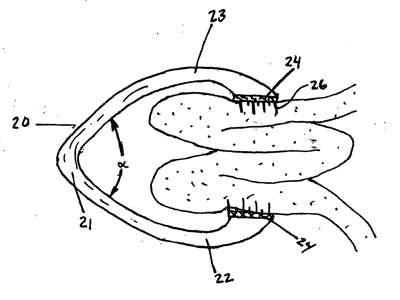
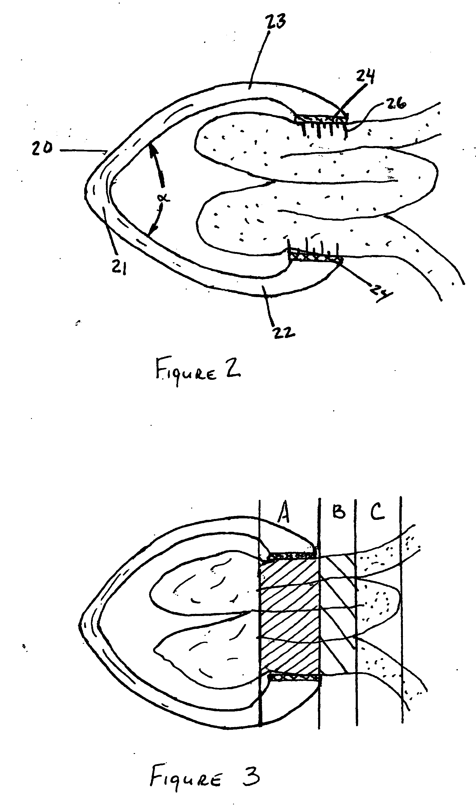
[0014] In another aspect of the present invention, the clamp also includes a cutting element that is located near the retention elements. The cutting element may be useful for gradually penetrating the tissue fold to facilitate severing the tissue fold once tissue necrosis has occurred. In still another aspect of the present invention, the clamp also includes a tethering element coupled to the clamp at one end. The tethering element is comprised of a cord with a tissue anchor coupled to the opposite end. The tissue anchor may be suitable for at least temporary coupling to a soft tissue wall at a location other than at the tissue fold. The tether may secure the clamp to the tissue wall so that the clamp can be retrieved later.

Problems solved by technology

Over time the clamping force may initiate mucosal layer pressure necrosis which can lead to an erosion or dissipation of the mucosal layer.

Method used


Image

  • Methods and apparatus for excising tissue and creating wall-to-wall adhesions from within an organ
  • Methods and apparatus for excising tissue and creating wall-to-wall adhesions from within an organ
  • Methods and apparatus for excising tissue and creating wall-to-wall adhesions from within an organ

Examples


Embodiment Construction

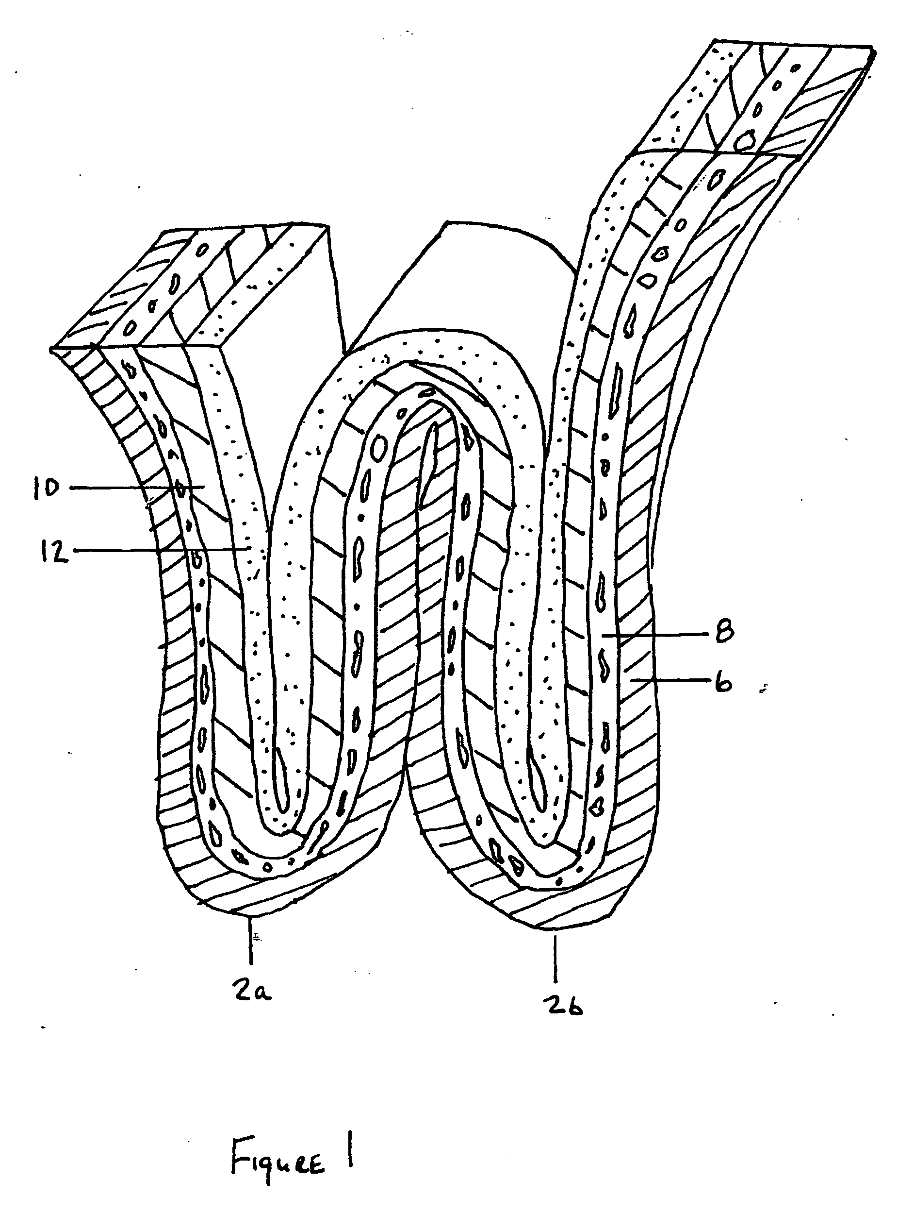
[0026] As has been described, folds of soft tissue are brought together and clamped for many reasons. As an example, folds of tissue are brought together for the purpose of reducing the volume of the stomach or other such organs. In the treatment of obesity, a gastric partition can be created in the stomach wall to restrict the intake of food to a smaller gastric volume. In this technique, the walls of the stomach are brought together using endoscopic techniques and secured together. When this procedure is performed using multiple sequential attachments, the volume of the stomach can be significantly reduced. In the first step of this procedure, the walls of the stomach are folded and usually two folds are brought into close proximity with each other as shown in FIG. 1. In this illustration, a section of a stomach wall 1 been formed by bringing together two folds 2a and 2b of stomach tissue together. The folds are brought together such that the inner surfaces of the stomach wall are...


PUM


Abstract

The preferred methods and devices described herein provide for creating adhesions between the tissue layers of folded soft tissue. The methods described can be accomplished with the use of an endoscope and once folds of tissue are formed and brought together using methods known in the art, the folds can be secured using a clamping system. This clamping system retains the tissue folds in a side by side relationship by exerting a clamping pressure against the folds. Over time the clamping pressure may initiate mucosal layer necrosis, deep tissue layer intermingling and tissue knitting.

Description

CLAIM OF PRIORITY [0001] This application claims priority under 35 U.S.C. § 119(e) to U.S. Provisional Application No. 60 / 679,284, filed May 9, 2005, the entire contents of which are hereby expressly incorporated by reference.BACKGROUND [0002] 1. Field of the Invention [0003] The present invention relates to devices and methods for joining segments of soft tissue together. [0004] 2. Description of the Related Art [0005] Often segments of soft tissue are brought together for the purpose of resecting tissue, providing anchors for other devices and for creating walls or partitions within an organ having a lumen. Sometimes a single wall of tissue is folded and brought together and other times two portions of soft tissue are grasped separately and then the two portions are brought into close proximity to each other and then joined together either permanently or temporarily. The joining of portions of soft tissue has traditionally been done using clamping, banding, suturing or stapling de...

Claims


Application Information

Patent Timeline
IPC IPC(8): A61B17/08
CPCA61B17/083A61B2017/2808A61B2017/081A61B17/085
Inventor KELLEHER, BRIANSWAIN, CHRISTOPHER PAUL
Owner KELLEHER BRIAN
Learn More