
Image-based artifact reduction in PET/CT imaging

a technology of image-based artifacts and pet/ct scans, applied in the field of medical imaging, can solve problems such as inability to accurately convert to mu map pixel values, difficulty in computing attenuation correction factors used, and inability to solve problems, etc., to achieve simple and robust effect of reducing errors in cardiac pet/

Inactive Publication Date: 2006-12-21
SIEMENS MEDICAL SOLUTIONS USA INC
View PDF2 Cites 38 Cited by
  • Summary
  • Abstract
  • Description
  • Claims
  • Application Information

AI Technical Summary

Benefits of technology

[0024] The present invention is provided for reducing errors in cardiac PET / CT in the case where an AICD is present in the patient's chest. Because the invention is simple and robust, it can be applied to other cases in which the ACF cannot be accurately derived from the CT images.
[0026] In another aspect of the present invention, the method provides for modifying the pixels in the CT image having a large HU value using a reassignment function of the original HU values that is continuous and smooth.
[0028] In still a further aspect of the invention, the method provides for after said identifying a region, morphologically dilating the region surrounding the pixels to enhance accuracy.

Problems solved by technology

In the field of combined Positron Emission Tomography and Computed Tomography (PET / CT), it is well known that difficulties are often encountered in computing the attenuation correction factors used.
Errors arise in the first step, in which the CT pixel values are incorrect, so that they cannot be converted accurately to mu map pixel values.
To date, this problem has not been resolved as a part of PET processing.
Specifically, this problem has not been resolved in the step of converting the pixel values to attenuation values.
However, it has been found that these methods do not work well when the metal moves during the measurement.
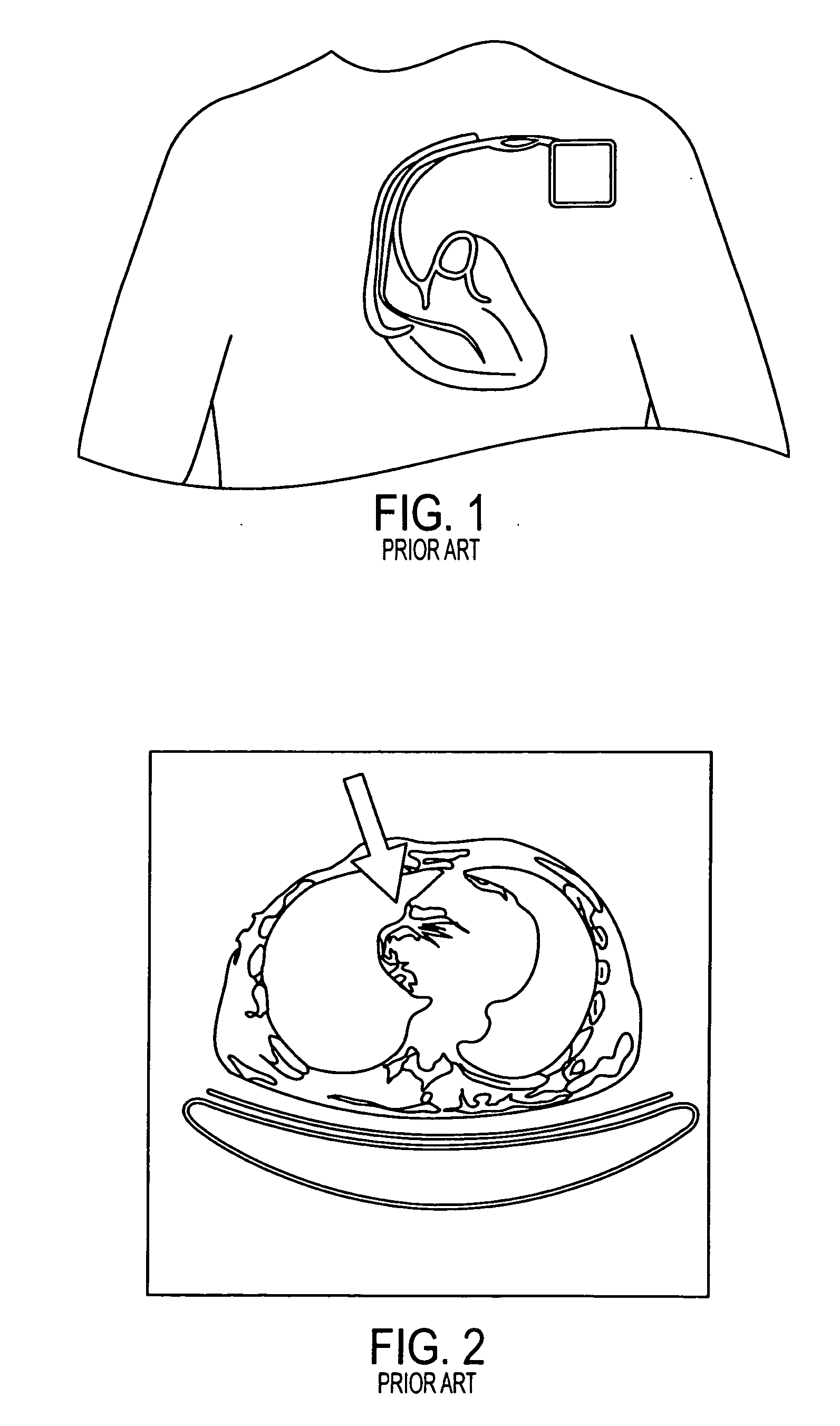
For CT machines, an AICD device presents more serious difficulties than a pacemaker.
When a CT machine reconstructs a section with the moving coil, the result is a metal artifact, with spurious high and low CT values in the region around the coil's actual location.
First, the CT image is an inaccurate picture of the anatomy.
Second, the PET portion of the PET / CT image may contain incorrect values.

Method used


Image

  • Image-based artifact reduction in PET/CT imaging
  • Image-based artifact reduction in PET/CT imaging
  • Image-based artifact reduction in PET/CT imaging

Examples


Embodiment Construction

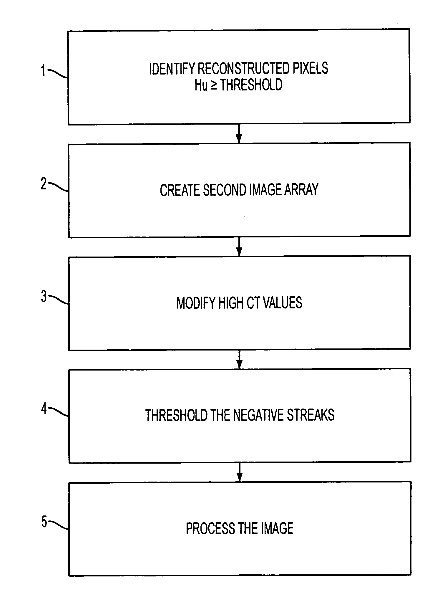
[0036] Exemplary embodiments of image-based artifact reduction (IBAR) methods for combined positron emission tomography and computed tomography (PET / CT) scans. The present embodiments are useful in the case in which the CT measurement of a PET / CT scan is corrupted by artifacts such as a moving piece of metal. The situation of reducing metal artifacts alone is known as metal artifact reduction, or MAR.

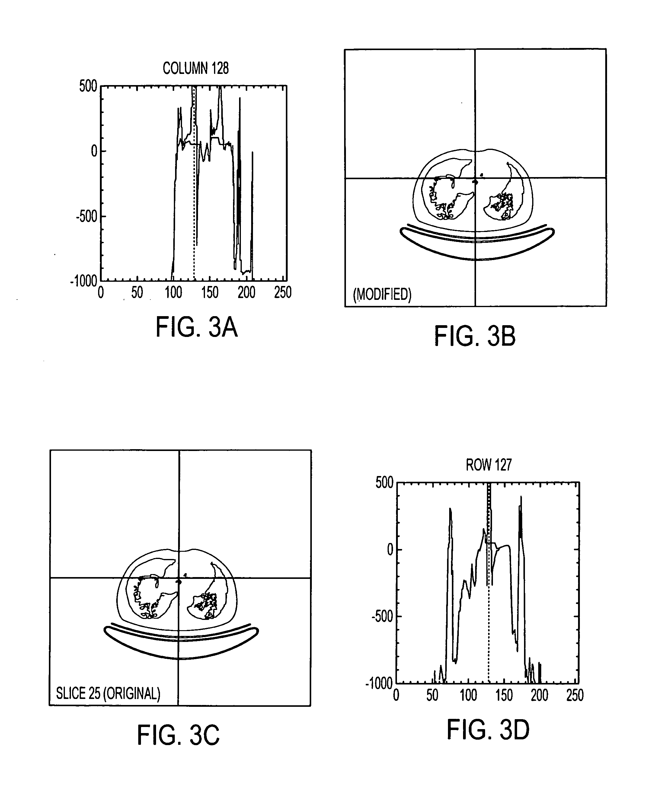
[0037] The exemplary IBAR method modifies the pixel values I(X,Y,Z) in a series of CT image slices. The image values are specified in Hounsfield units (HU). Properly functioning CT equipment creates images in which water is assigned a value of zero (0 HU), air is assigned a value of approximately −1000 HU, and bone and metal are assigned values greater than zero (0 HU). In the PET / CT processing software that has been used, images are first reduced from arrays of size 512×512 pixels to size 256×256 with a rebinning procedure, in which groups of four pixels in the 512×512 matrices are av...


PUM


Abstract

A method for reducing image-based artifacts in combined positron emission tomography and computed tomography (PET / CT) scans. The method includes identifying pixels in a CT image having a large HU value, identifying a region surrounding the pixels, and modifying a value of each pixel within the region.

Description

CROSS-REFERENCE TO RELATED APPLICATIONS [0001] This application claims priority from U.S. Provisional Ser. No. 60 / 691,811 titled “Image Based Artifact Reduction In PET / CT Imaging” filed on Jun. 17, 2005, the entire contents of which is incorporated herein by reference.BACKGROUND OF THE INVENTION [0002] 1. Field of Invention [0003] The present invention pertains to the field of medical imaging using combined Positron Emission Tomography (PET) and Computed Tomography (CT) modalities. More particularly, this invention is directed toward a method for reducing image-based artifacts in a PET / CT scan. [0004] 2. Description of the Related Art [0005] In the field of combined Positron Emission Tomography and Computed Tomography (PET / CT), it is well known that difficulties are often encountered in computing the attenuation correction factors used. Normally this computation is performed as a digital calculation in computers used for PET / CT. The typical procedure for deriving attenuation correct...

Claims


Application Information

Patent Timeline
Patent Type & Authority Applications(United States)
IPC IPC(8): G06K9/00
CPCG06T11/008G06T5/002G06T5/005G06T5/30G06T2207/30048G06T2207/10081G06T2207/10104G06T2207/20032G06T2200/04A61B6/5258G06T5/77G06T5/70
Inventor HAMILL, JAMES J.FAUL, DAVID D.
Owner SIEMENS MEDICAL SOLUTIONS USA INC
Learn More