Methods for purifying protein

a protein and purification technology, applied in the field of protein purification, can solve the problem that other protein contaminants may also be present in a sampl

Inactive Publication Date: 2007-01-11
IMMUNEX CORP
View PDF12 Cites 5 Cited by
  • Summary
  • Abstract
  • Description
  • Claims
  • Application Information

AI Technical Summary

Problems solved by technology

However, if the proteins are manufactured for therapeutic use, the presence of other proteins, including a protein used as part of an affinity adsorbent, which can leach into a sample during affinity chromatography, is of concern.
In addition, other protein contaminants may also be present in a sample, such as, for example, proteins derived from host cells that produce the protein being purified.

Method used


Image

  • Methods for purifying protein
  • Methods for purifying protein

Examples


example 1

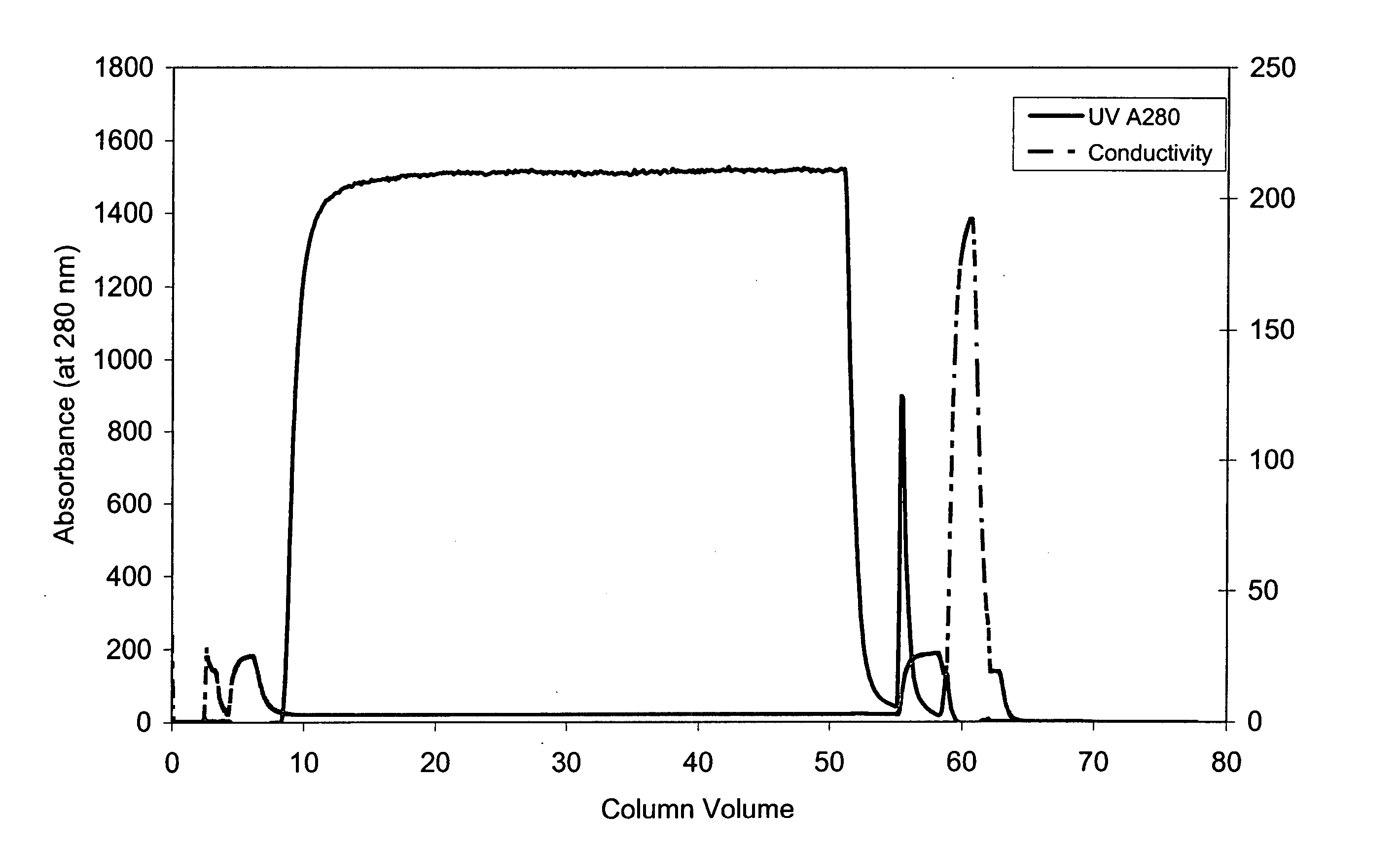
Reduction in Levels of Protein A in a Sample Comprising TNFR:FC Using Hydroxyapatite Chromatography

[0068] This experiment demonstrates that hydroxyapatite chromatography can reduce levels of residual protein A in a protein sample comprising TNFR:FC that contains a defined amount of protein A, which can form a complex with TNFR:FC.

[0069] A column of ceramic hydroxyapatite (Type II, Bio-Rad, 80μ) 18 cm in height and 1.6 cm in internal diameter was pre-equilibrated with two column volumes of 0.4 molar sodium phosphate, pH 6.8 and equilibrated with four column volumes of 25 millimolar sodium phosphate, pH 6.8, which has a conductivity of 2.8 milliSiemens (mS). A protein sample (5.11 milligrams / milliliter) in 668 milliliters of 25 mM sodium phosphate, pH 6.8 comprising TNFR:FC and protein A (209 ppm) was loaded onto the column. The amount of protein A in the sample was determined using an ELISA assay. The flow-through liquid, containing TNFR:FC, was collected. The column was washed wit...

example 2

Reduction in the Levels of Host Cell Proteins in a Sample Comprising TNFR:FC Using Hydroxyapatite Chromatography

[0070] The following experiment demonstrates that the levels of host cell proteins in a sample comprising TNFR:FC can be reduced by hydroxyapatite chromatography.

[0071] A column of ceramic hydroxyapatite (Type II, Bio-Rad, 80μ) 10 cm in height and 1.1 cm in internal diameter was pre-equilibrated with two column volumes of 0.3 molar sodium phosphate, pH 6.8 and equilibrated with three column volumes of 25 millimolar sodium phosphate, pH 6.8, which has a conductivity of 2.8 mS. About 1.9 grams of protein in a volume of 382 milliliters of 25 millimolar sodium phosphate, pH 6.8 was loaded onto the column. This sample included TNFR:FC, which comprised the vast majority of the sample, and host cell proteins (545 ppm). The amount of host cell proteins in the sample was determined using ELISA assays. The flow-through liquid, comprising TNFR:FC, was collected. The column was wash...

example 3

Reduction in Residual Protein A and Other Proteins in a Sample of TNFR:FC by Ceramic Hydroxyapatite Chromatography

[0073] The following experiment shows that levels of protein A and other protein contaminants can be simultaneously reduced using hydroxyapatite chromatography.

[0074] A column of ceramic hydroxyapatite (Type II, Bio-Rad, 80μ) 18 cm in height and 1.6 cm in internal diameter was pre-equilibrated with two column volumes of 0.4 M sodium phosphate, pH 6.8 and equilibrated with three column volumes of 25 millimolar sodium phosphate, pH 6.8. About 3.2 grams of protein at about 5 mg / ml in 25 millimolar sodium phosphate comprising TNFR:FC, Protein A, which forms a complex with TNFR:FC under these conditions, (between about 97 ppm and about 106 ppm), and other process related impurities (PRI; between about 59 ppm and about 67 ppm) was loaded onto the column. The amounts of Protein A and PRI in the sample were determined using ELISA assays. The flow-through liquid, containing TNF...


PUM

PropertyMeasurementUnit
pHaaaaaaaaaa
molecular weightaaaaaaaaaa
heightaaaaaaaaaa

Abstract

A method is provided for separating a protein from one or more other proteins using hydroxyapatite chromatography in which the protein does not bind to hydroxyapatite but the other protein(s) does. In some embodiments, a second protein affixed to a solid support has been used previously to purify the protein by affinity chromatography, and small amounts of the second protein are introduced in the sample during this process. The protein being purified can comprise at least one constant antibody immunoglobulin domain. The second protein can bind to proteins comprising such a domain.

Description

[0001] This application is a continuation of U.S. application Ser. No. 10 / 327,495, filed Dec. 20, 2002, the disclosure of which is incorporated by reference herein, which claims benefit of three provisional applications, U.S. Application No. 60 / 343,363, filed Dec. 21, 2001, U.S. Application No. 60 / 347,189, filed Jan. 8, 2002, and U.S. Application No. 60 / 364,272, filed Mar. 12, 2002, the disclosures of which are incorporated by reference herein.FIELD OF THE INVENTION [0002] The invention relates to protein purification, in particular, purification utilizing hydroxyapatite chromatography. BACKGROUND [0003] Proteins A and G are often employed to purify antibodies by affinity chromatography. See e.g. R. Vola et al. (1994), Cell Biophys. 24-25: 27-36; Aybay and Imir (2000), J. Immunol. Methods 233(1-2): 77-81; Ford et al. (2001), J. Chromatogr. B 754: 427-435. These proteins are useful because they bind to a constant (FC) portion of many different antibodies. Recombinant fusion proteins ...

Claims


Application Information

Patent Timeline
Patent Type & Authority Applications(United States)
IPC IPC(8): C07K16/46C12N15/09C07K1/16C07K1/20C07K1/22C07K14/705C07K14/715C07K16/18C07K19/00C12P21/02C12P21/08
CPCC07K1/16C07K1/22C07K2319/30C07K14/7151C07K2319/00C07K14/70575
Inventor VEDANTHAM, GANESHBROOKS, CLAYTON A. IIIREEDER, JOANNE M.GOETZE, ANDREW M.
Owner IMMUNEX CORP
Why Eureka
Try Eureka