
Method for locating brain lesion

a brain lesion and brain tissue technology, applied in the field of brain lesion locating methods, can solve the problems of time-consuming stereotaxi, doubling the operating room time, and not being user-friendly,

Inactive Publication Date: 2007-01-18
ST BARNABAS HEALTH CARE SYST
View PDF1 Cites 6 Cited by
  • Summary
  • Abstract
  • Description
  • Claims
  • Application Information

AI Technical Summary

Problems solved by technology

Stereotaxi is therefore time-consuming, often doubling the operating room time.
In addition, the equipment and systems used to perform stereotaxi are cumbersome, not user friendly, and quite expensive.

Method used


Image

  • Method for locating brain lesion
  • Method for locating brain lesion
  • Method for locating brain lesion

Examples


Embodiment Construction

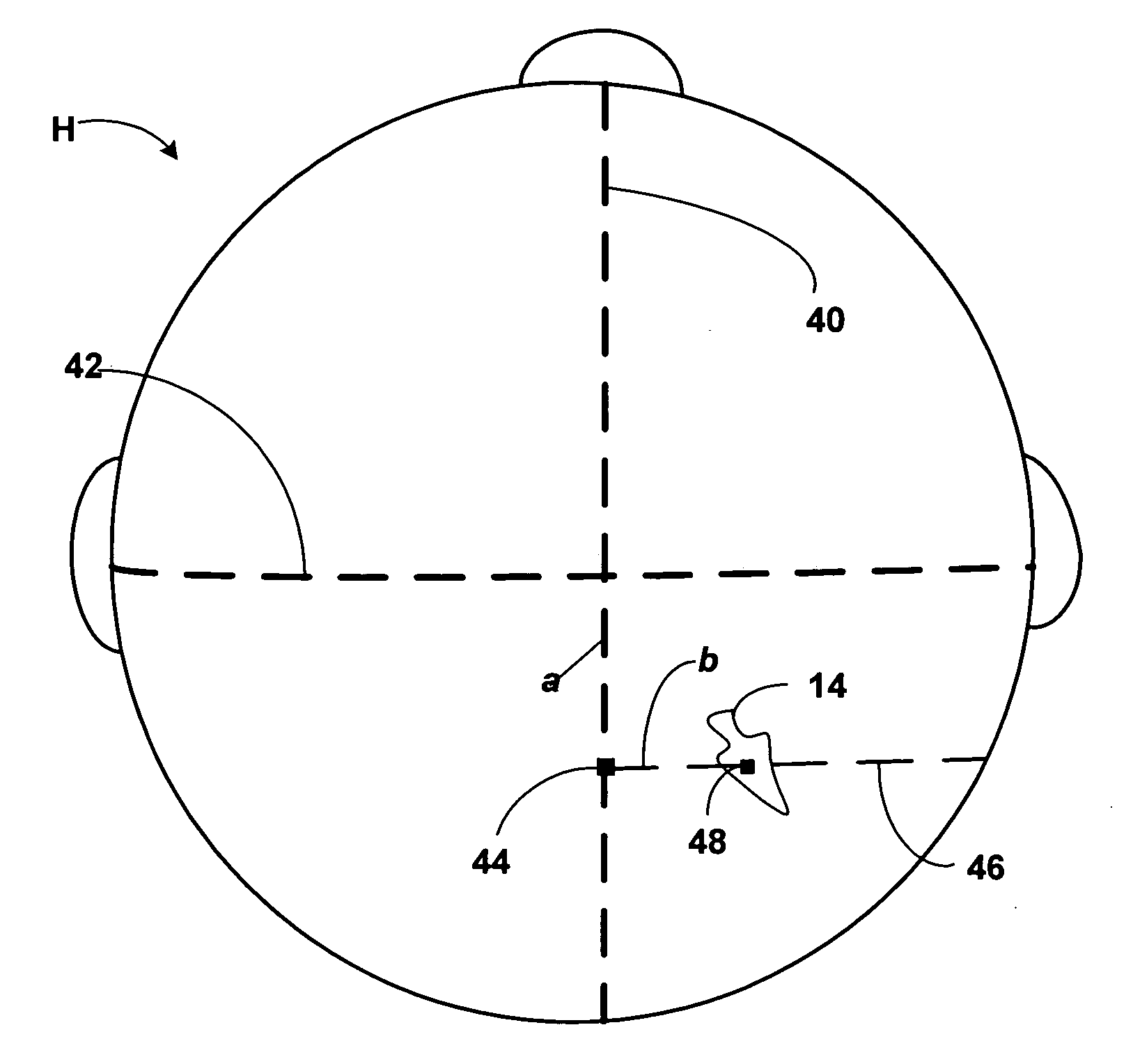
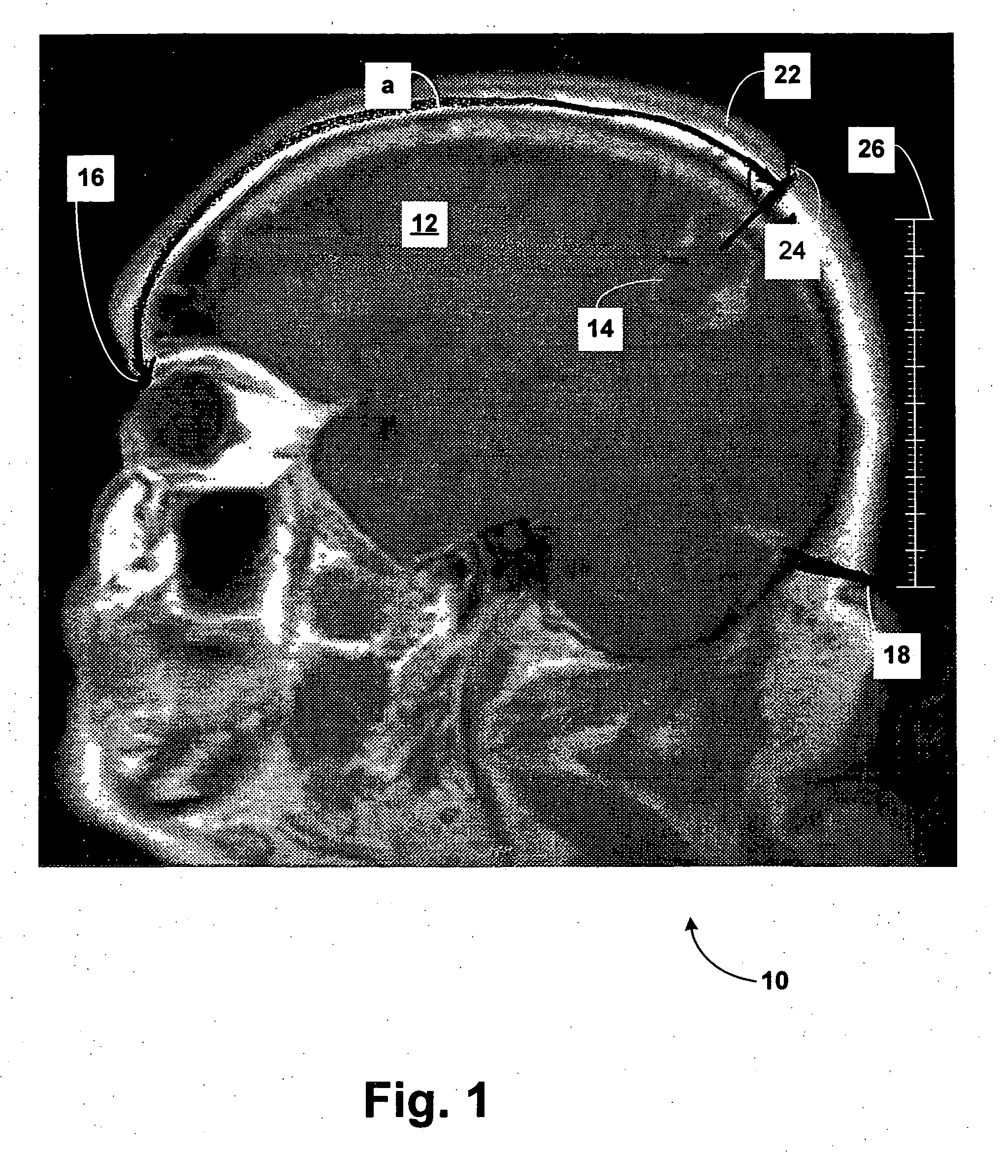
[0012] As explained above, a surgeon utilizes a brain scan image to locate a brain lesion and to plan the operation to treat it. In the operating room, he transfers distance measurements derived directly from the brain scan onto the patient's cranium, thereby establishing the appropriate location and orientation of the bone flap.

[0013]FIG. 1 is a saggital MRI 10 of a patient's head showing the brain which has a lesion 14. The figure also shows the patient's nasion 16 (the recess above the nose) and inion 18 (the recess at the base of the skull). As may be seen, MRI 10 has a scale 26, which is typical. The scale 26 may be in the centimeters (large gradations), indicating the length of a centimeter on the patient's head.

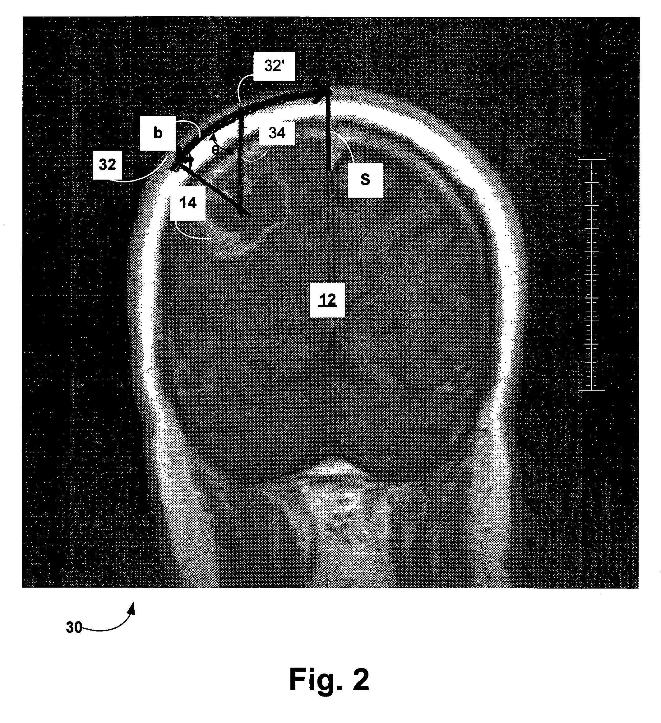
[0014] As an initial step, the surgeon measures along the surface of the cranium 22 the distance a, the distance measured on the MRI from the nasion 16 to a point overlying the center of the lesion 14. This measurement ends at the point 24. The depth of the lesion in...


PUM


Abstract

A surgeon utilizes a brain scan image to locate a brain lesion and to plan the operation to treat it. Distance measurements representing the location of the lesion are derived from various views of a brain scan image. In the operating room, he transfers these distance measurements onto the patient's cranium in a “warped” or curvilinear Cartesian coordinate system defined by intersecting orthogonal lines extending along the surface of the patient's cranium. As a result, the lesion is localized and the surgeon may establish the appropriate location and orientation of a bone flap to be used to access the lesion.

Description

BACKGROUND OF THE INVENTION [0001] In performing cranial surgery, it is the common practice to form a bone flap in the patient's cranium so as to gain access to the brain. While standard bone flaps may be used during neurosurgery for specific lesions, such as for an aneurysm clipping and certain types of skull base surgery procedures, removal of most cranial lesions requires a specific “customized” flap. Requirements of present day neurosurgery make it imperative that the flap be accurate and as small as possible. This is typically achieved by frame-based or frameless stereotaxi. [0002] Frame-based stereotaxi involves the placement of a frame on the patient's head, which provides a spatial frame of reference and the acquisition of a brain scan image, such as a Computed Tomography (CT) scan or Magnetic Resonance imaging (MRI). Frameless stereotaxi requires the placement of fiducial markers on the patient's head. Another scan is necessary prior to any surgical procedure, which require...

Claims


Application Information

Patent Timeline
IPC IPC(8): G06K9/00A61B5/05
CPCA61B5/055A61B5/1075A61B19/52A61B19/20A61B6/03A61B90/10A61B90/36A61B5/05
Inventor HUBSCHMANN, OTAKAR R.
Owner ST BARNABAS HEALTH CARE SYST
Learn More