
Method and apparatus for predicting structure of transmembrane proteins
a transmembrane protein and structure technology, applied in the field of three-dimensional structure prediction of proteins, can solve the problems of time-consuming and expensive techniques, difficult to determine the tertiary structure of proteins, and much effort put into elucidation, and achieve the effect of fast and accurate procedures
- Summary
- Abstract
- Description
- Claims
- Application Information
AI Technical Summary
Benefits of technology
Problems solved by technology
Method used
Image

Examples
example 1
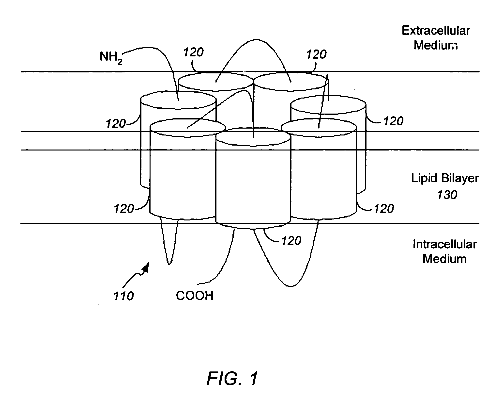
[0038] The protocol described above was tested on bacteriorhodopsin (BRDP), a membrane protein for which the crystal structure has been fitted with fair accuracy in the transmembrane region of the protein. Starting from the sequence of bacteriorhodopsin, and without using coordinates from the crystal structure, the protocol described above was used to build the complete protein model. The membrane was represented by bilayers of diphosphatidyl glycerophosphate that is the lipid present in the purple membrane from Halobacterium halobium. Although the sequence homology between BRDP and the ORs is not high (less than 30%), they share the same tertiary motif common to α-helical transmembrane proteins: a 7-helix barrel.
[0039] As shown in FIG. 5, the predicted tertiary structure for BDRP compares favorably with the known crystal structure. The overall rms deviation in coordinates of Cα atoms from the crystal structure for the final model is 5.98 Å for all 221 aa. The overall rms deviation...
example 2
[0040] Modeling of six olfactory receptors. Sequences for ORS25, ORS18, ORS19, ORS6, ORS46 and ORS50 were obtained from taken from Malnic et al. For each receptor, the membrane was simulated by using explicit lipid bilayers of dilauroylphosphatidyl choline. The choice of lipid in the OR case is supported by experimental indications, Gimenez, C. (1998) Rev. Neurol. (Paris) 26, 232-239; Kiefer, H. et al. (1996) Biochemistry 35, 16077-16084, that the membrane surrounding the ORs in vivo can be satisfactorily simulated by using a single-component lipid system of dilauroylphosphatidyl choline. Final atomic level models for the six receptors are shown in FIG. 6. Predicted structural models for S6, S18, S19, S25, S46 and S50 are included in PDB format in Tables 2 through 7, respectively, submitted on compact disc and incorporated by reference above. An explanation of the PDB file format can be found at http: / / www.rcsb.org / pdb / . See also Berman, H. M., et al. (2000) Nucleic Acids Res. 28, 2...
PUM
| Property | Measurement | Unit |
|---|---|---|
| time | aaaaa | aaaaa |
| dielectric constant | aaaaa | aaaaa |
| structure | aaaaa | aaaaa |
Abstract
Description
Claims
Application Information