Systems and methods for predicting the structure and function of multipass transmembrane proteins

a multi-pass transmembrane protein and protein technology, applied in the field of systems and methods for predicting the structure and function of multi-pass transmembrane proteins, can solve the problems of undesirable side effects, time-consuming and expensive techniques, and difficult to determine the tertiary structure of proteins

Inactive Publication Date: 2005-06-23
CALIFORNIA INST OF TECH
View PDF3 Cites 21 Cited by
  • Summary
  • Abstract
  • Description
  • Claims
  • Application Information

AI Technical Summary

Benefits of technology

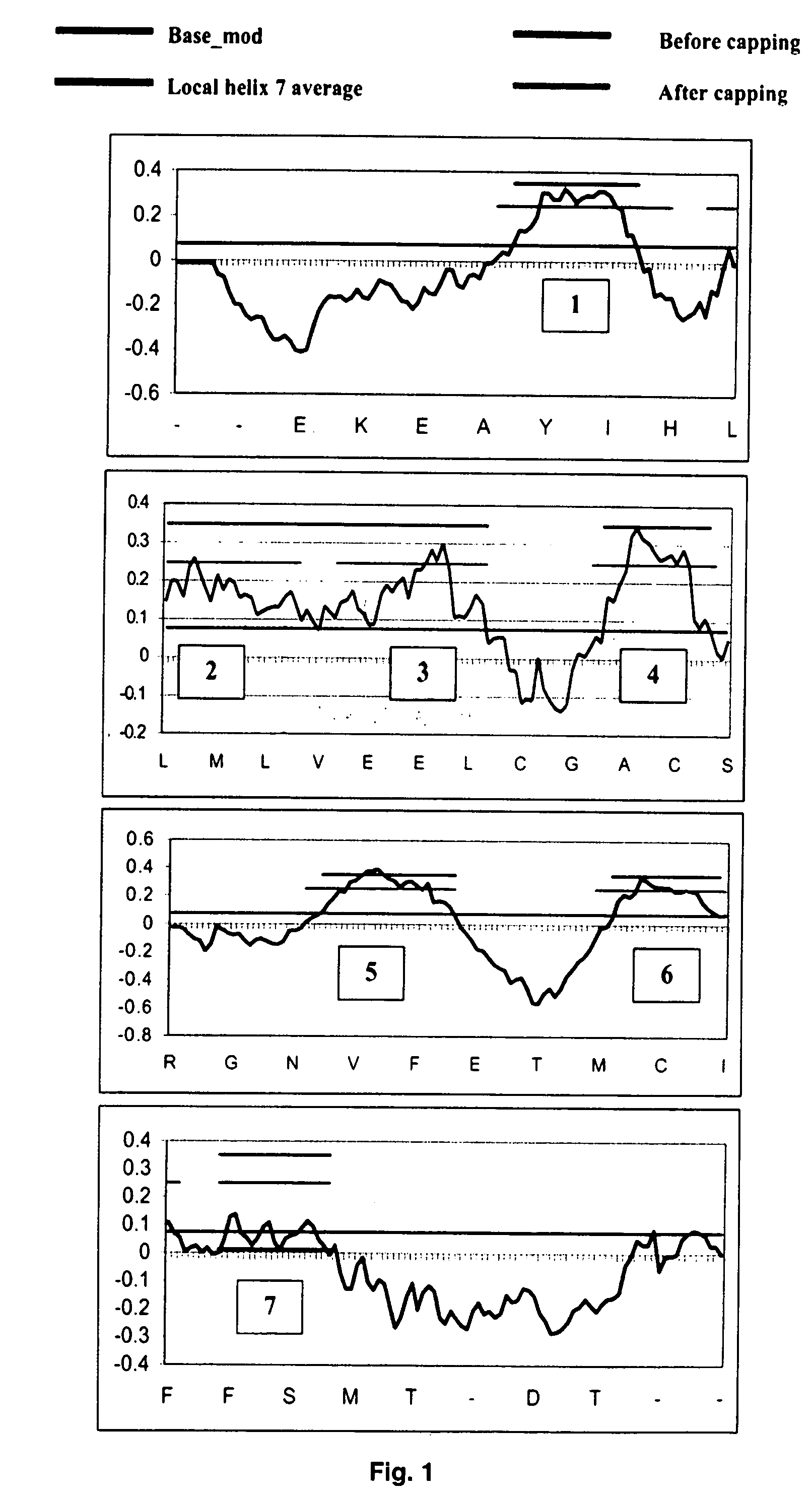
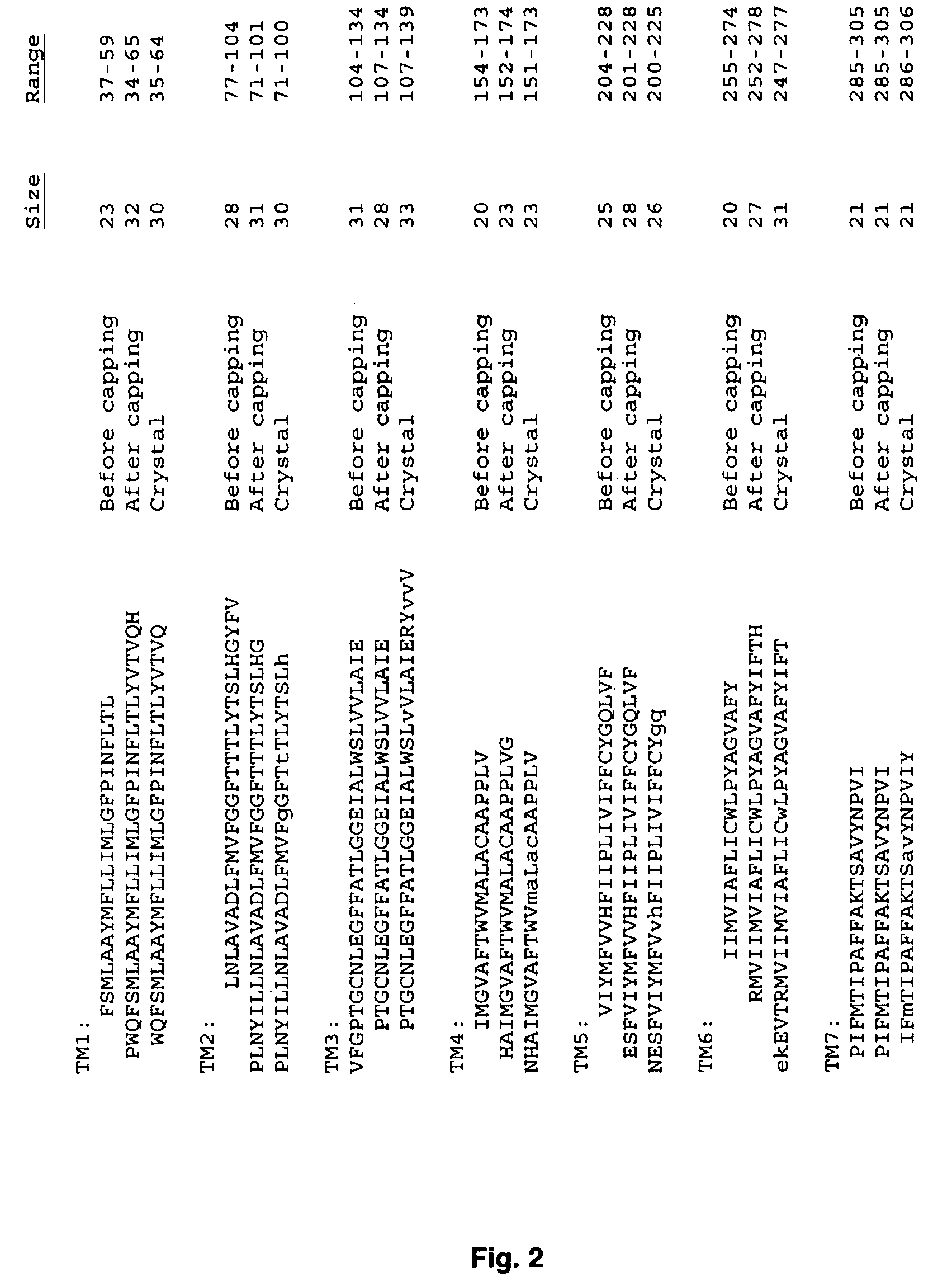
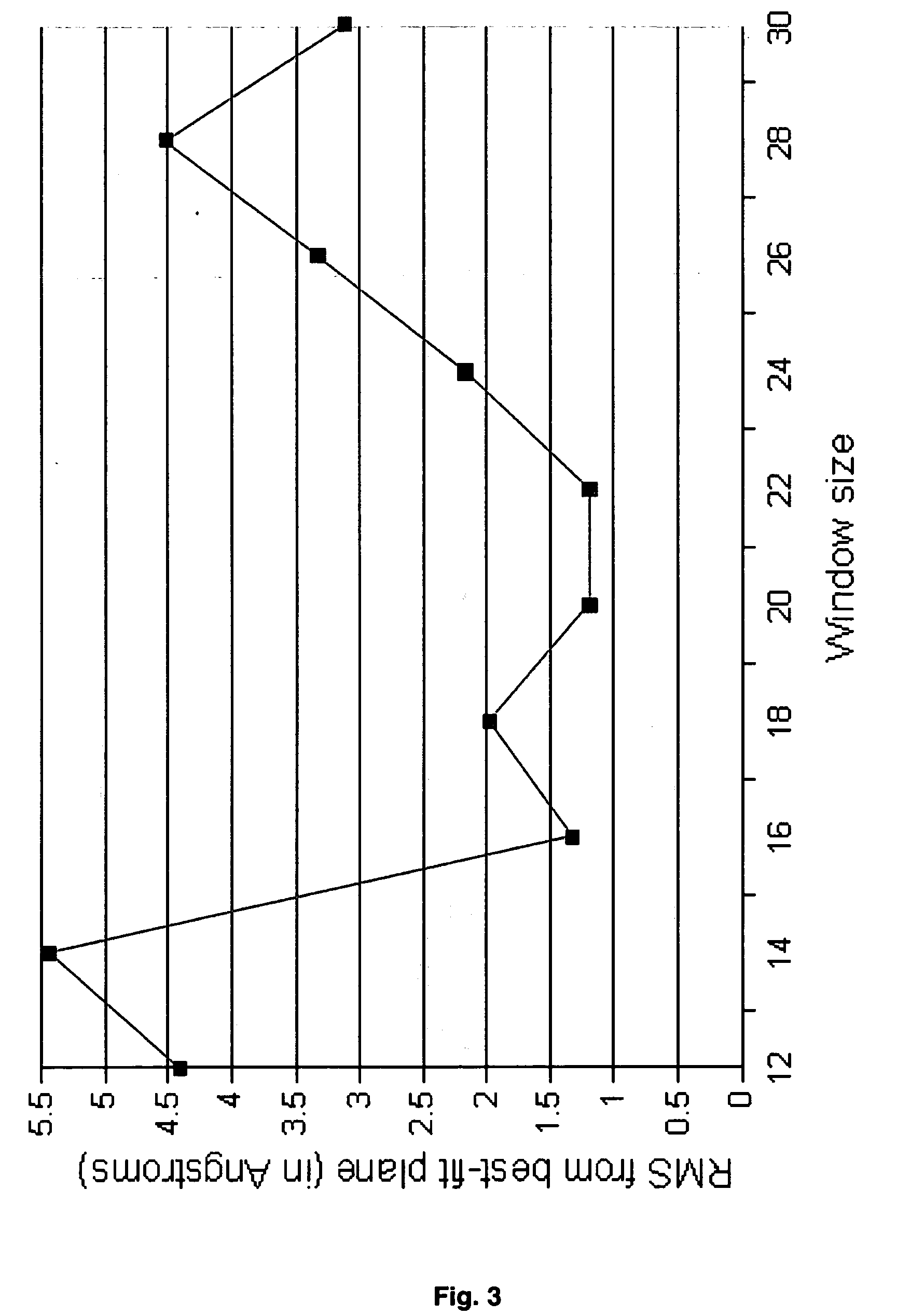
[0015] The invention provides a hierarchical protocol using multiscale molecular dynamics and molecular modeling methods to predict the structure of multipass membrane proteins, such as G-Protein Coupled Receptors (GPCRs), from primary amino acid sequence of a target protein. The protocol features a combination of coarse grain sampling methods, such as hydrophobicity analysis, followed by coarse grain molecular dynamics and atomic level molecular dynamics, including accurate continuum solvation. The methods also include energy optimization to determine the rotation of helices in the (seven-helical) TM bundle, and optimization of the helix translations along their axes and rotational optimization using hydrophobic moment of the helices, to provide a fast and accurate procedure for predicting tertiary structure.
[0055] In one embodiment, for each of the at least one helix, the RotMin comprises: (a) designating one of said at least one helix as active helix; (b) keeping the main chain of said active helix rigid while rotating said main-chain for a grid of rotation angles; (c) optimizing side-chain positions of all residues for all helices in said TM bundle; (d) minimizing energy for said active helix in the field of all other helices; (e) repeating (a)-(d) for each of said at least one helix.
[0063] In one embodiment, for each individual TM region, step (3) is effectuated by: (a) placing all side-chains; (b) minimizing energy using molecular dynamics (MD).
[0065] In one embodiment, step (b) is effectuated by simulations at 300 K for 500 ps, and minimizing the structure with the lowest potential energy in the last 250 ps using conjugate gradients.

Problems solved by technology

Determining a protein's tertiary structure is more difficult.
However, these techniques can be time consuming and expensive, and not all proteins are equally amenable to structural examination by these methods.
In addition, many different types of GPCRs are similar enough that they are affected by the antagonists or agonists for other types (e.g., among adrenergic, dopamine, serotonin, and histamine receptors), leading often to undesirable side effects.
This makes it difficult to develop drugs to a particular subtype without side effects resulting from cross-reactivity to other subtypes.
However, although much effort has been put into elucidating the structure of GPCRs, only a very small number of complete 3D structures of transmembrane proteins are known from experiments (e.g., bacteriorhodopsin and bovine rhodopsin).
In fact, there is no atomic-level structure available for any human GPCRs.
Consequently, design of subtype-specific drugs for GPCR targets is a very tedious empirical process, often leading to drugs with undesirable side effects.
The difficulty in obtaining three-dimensional structures for GPCRs is obtaining high-quality crystals of these membrane-bound proteins sufficient to obtain high-resolution x-ray diffraction data, and the difficulty of using NMR to determine structure on such membrane-bound systems.
For globular proteins, there have been significant advances in predicting the three-dimensional structures by using sequence homologies to families of known structures (Marti-Renom et al., 2000); however, this is not practical for GPCRs, inasmuch as a high-resolution crystal structure is available for only one GPCR, bovine rhodopsin-which has low homology (<35%) to most GPCRs of pharmacological interest.
Such validation methods are also lacking in the art.

Method used


Image

  • Systems and methods for predicting the structure and function of multipass transmembrane proteins
  • Systems and methods for predicting the structure and function of multipass transmembrane proteins
  • Systems and methods for predicting the structure and function of multipass transmembrane proteins

Examples


example 1

[0380] Validation of the Force Fields

[0381] The crystal structure of bovine rhodopsin (resolution, 2.80 Å) was downloaded from the protein structure database (PDB entry 1F88). The Hg ions, sugars, and waters were deleted from this structure. This crystal structure is missing 10 complete residues in loop regions and the side-chain atoms for 15 additional residues. We added the missing residues and side chains using WHATIF (Vriend, 1990). Then we added hydrogens to all the residues using the PolyGraf software. We then fixed the TM helices and minimized (using conjugate gradients) the structure of the loop region to a root mean-square force of 0.1 kcal / mol per Å. The potential energy of the entire structure of rhodopsin was then minimized (using conjugate gradients) to a root mean-square force of 0.1 kcal / mol per Å. This minimized structure deviates from the x-ray crystal structure by 0.29 Å coordinate root mean-square (CRMS) error over all atoms in the crystal structure. This is with...

example 2

[0382] Various Bovine Rhodopsin Structures (Crystal or Predicted)

[0383] The crystal structure for the retinal / rhodopsin complex has a well-defined β-sheet structure for EC-II, which might be involved as a mobile gate for entry of 11-cis-retinal on the extracellular side of rhodopsin. Such a gating mechanism is illustrated in FIG. 4, in which the helix 3 coupled to this loop by a cysteine bond is the gatekeeper which responds to signaling structural substrates of rhodopsin as follows:

[0384] When rhodopsin binds 11-cis-retinal, the ground state conformation of the receptor is stabilized, thus shifting helix 3 toward the intracellular side (forming the D(E)RY-associated salt bridges at that end) and closing the EC-II loop. In fact, 11-cis-retinal has been shown to be an inverse agonist for G-protein signaling (Okada et al., 2001).

[0385] In response to absorption of a photon, the 11-cis-retinal isomerizes to the all-trans conformation, inducing helix 3 to shift toward the extracellul...

example 3

Validation of the HierDock Protocol on the Crystal Structure of Bovine Rhodopsin

[0397] Bovine rhodopsin (a member of the opsin family) is the only GPCR to be crystallized in its entirety at a high resolution (2.8 A). Thus we used this system as a test to validate the HierDock protocol for predicting the binding sites of GPCRs.

[0398] To test HierDock, we used the Apo / closed(xtal) structure with the retinal removed and minimized. First we did a complete HierDock scan as outlined above to predict the binding of 11-cis-retinal to bovine rhodopsin. The crystal structure of rhodopsin has the 11-cis-retinal covalently bound to Lys-296 (between the aldehyde of 11-cis-retinal and the N of the Lys), but for docking we cannot have a covalent bond to the crystal. Thus we docked the full 11-cis-retinal ligand (containing a full aldehyde group) and considered the Lys-296 to be protonated.

[0399] We applied Steps 1-2 of the HierDock described above for all 13 overlapping regions for Step 2 show...


PUM

PropertyMeasurementUnit
rotation anglesaaaaaaaaaa

Abstract

The invention provides computer-implemented methods and apparatus implementing a hierarchical protocol using multiscale molecular dynamics and molecular modeling methods to predict the structure of transmembrane proteins such as G-Protein Coupled Receptors (GPCR), and protein structural models generated according to the protocol. The protocol features a combination of coarse grain sampling methods, such as hydrophobicity analysis, followed by coarse grain molecular dynamics and atomic level molecular dynamics, including accurate continuum solvation. Also included are energy optimization to determine the rotation of helices in the (seven-helical) TM bundle, and optimization of the helix translations along their axes and rotational optimization using hydrophobic moment of the helices, to provide a fast and accurate procedure for predicting GPCR tertiary structure.

Description

REFERENCE TO RELATED APPLICATIONS [0001] This application claims the benefit of the filing date, under 35 U.S.C. 119(e), of U.S. Provisional Application No. 60 / 494,971, filed on Aug. 13, 2003, the entire contents of which is incorporated herein by reference.GOVERNMENT SUPPORT [0002] The invention described herein was supported, in whole or in part, by Grant Nos. NIHBRGR01-GM625523, NIH-R29AI40567, NIH-HD36385, R01GM6225301, and other grants such as DAAG55-98-1-0266, from other U.S. Government Agencies including DOE (ASCI ASAP) and NSF. The U.S. Government has certain rights in this invention.BACKGROUND OF THE INVENTION [0003] Proteins are linear polymers made up of 20 different naturally-occurring amino acids. The particular linear sequence of amino acid residues in a protein is said to define the protein's primary structure. In its natural environment, a protein folds into a three-dimensional structure determined by its primary structure, and by the chemical and electronic interact...

Claims


Application Information

Patent Timeline
Patent Type & Authority Applications(United States)
IPC IPC(8): G16B15/20G01N33/48G01N33/50G01N33/53G16B30/10G16B30/20
CPCG06F19/22G06F19/16G16B15/00G16B30/00G16B15/20G16B30/10G16B30/20
Inventor TRABANINO, RENEVAIDEHI, NAGARAJANHALL, SPENCERGODDARD, WILLIAMFLORIANO, WELY
Owner CALIFORNIA INST OF TECH
Why Eureka
Try Eureka