
Method for forming an endoscope articulation joint

a technology of articulation joints and endoscopes, applied in the field of medical devices, can solve the problems of limited flexibility, limited column strength, limited operator control of articulation joints along endoscope length, etc., and achieve the effect of low cost and easy mass production

Active Publication Date: 2007-03-01
BOSTON SCI SCIMED INC
View PDF99 Cites 155 Cited by
  • Summary
  • Abstract
  • Description
  • Claims
  • Application Information

AI Technical Summary

Benefits of technology

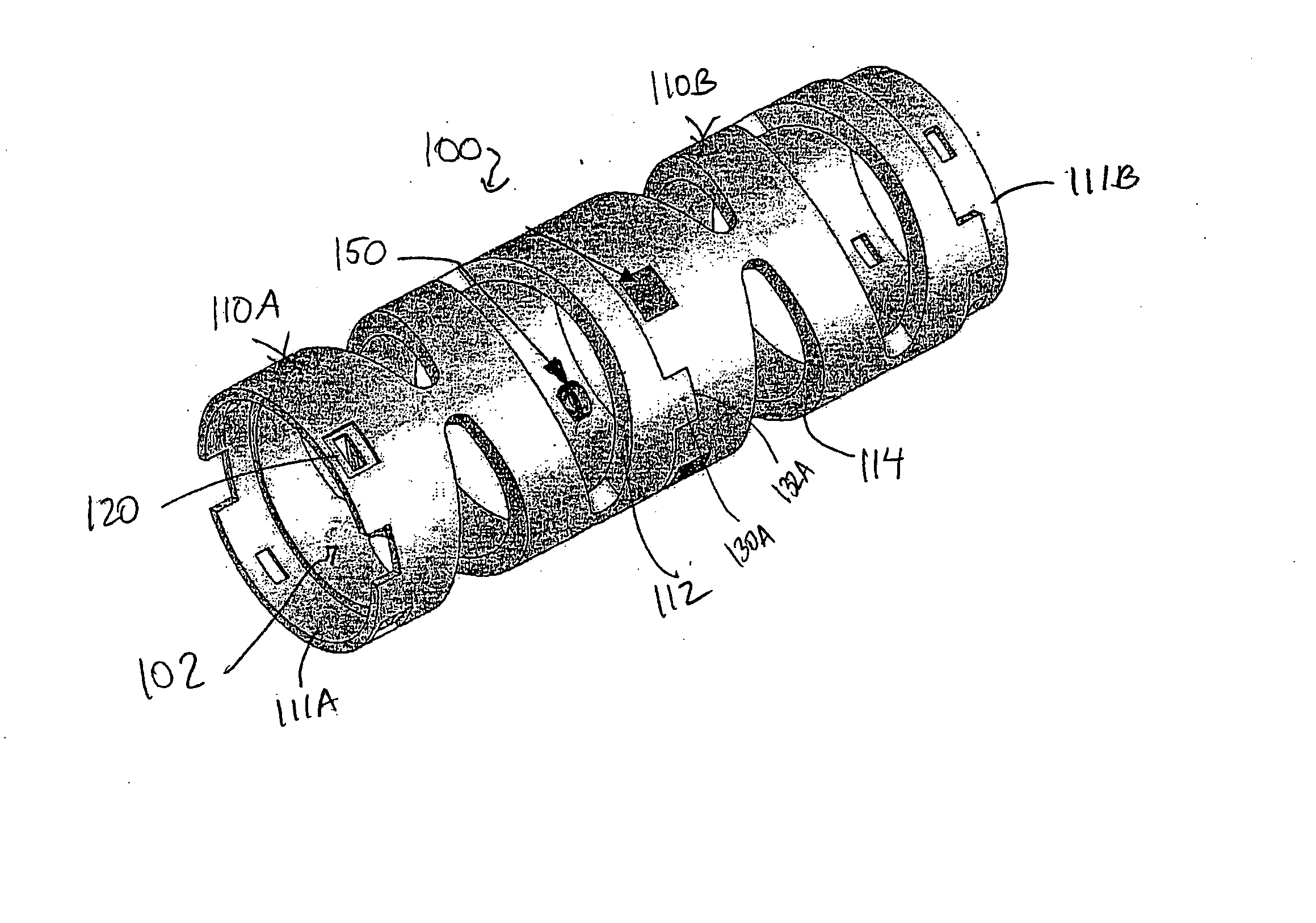
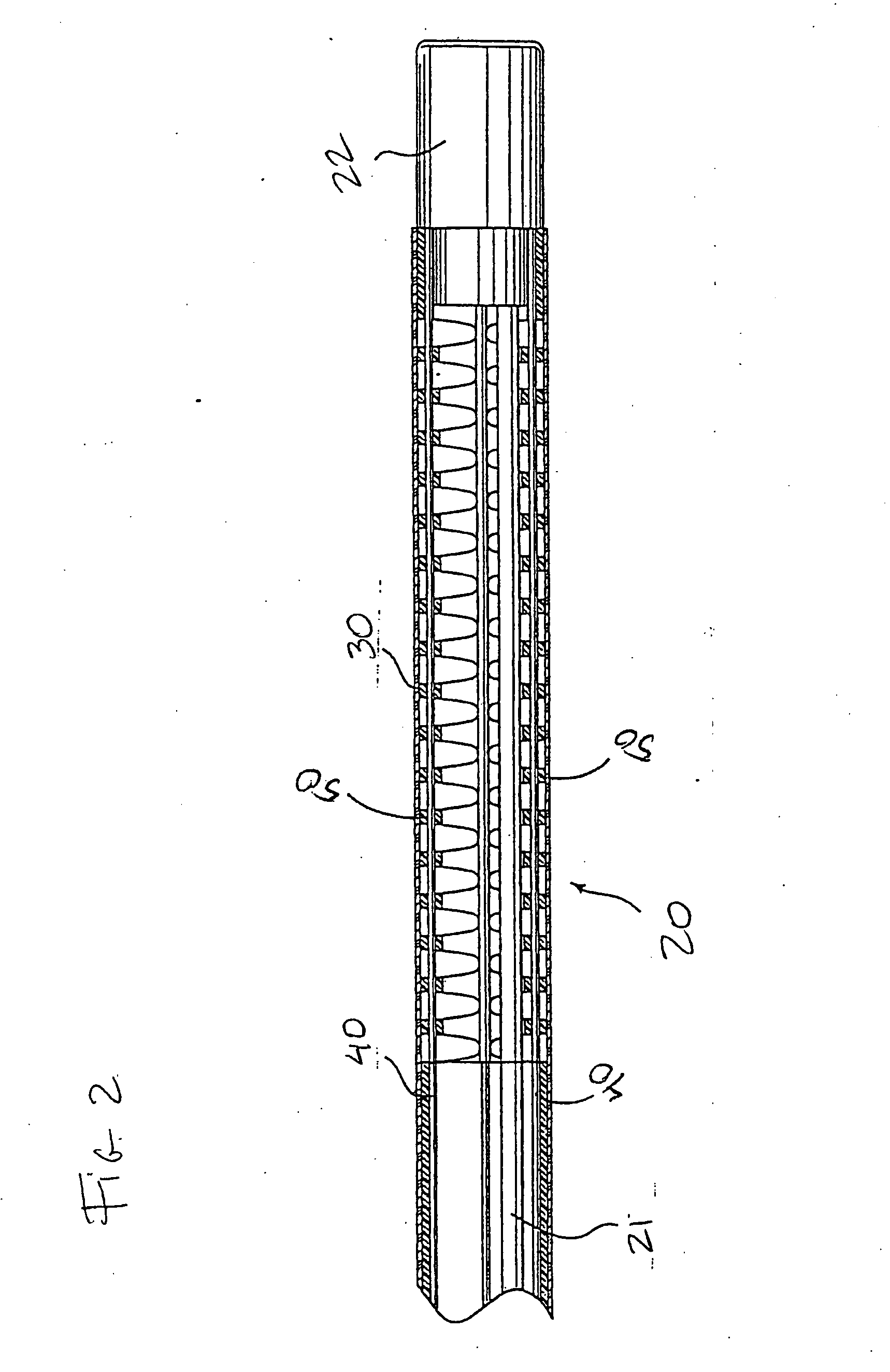
[0007] To address these and other concerns, in one embodiment the present invention is an articulation joint for use in a medical device such as an endoscope. The articulation joint is comprised of a number of low cost, easily mass produced components that allow the distal end of the endoscope to be bent in a desired direction by one or more control cables. In one embodiment, the articulation joint body comprises a plurality of interconnecting segments. Each segment comprises a cylinder with an outer wall and a central lumen. The outer wall includes a number of hinge elements therein and a series of slots therethrough. A plurality of cable guide elements having a passage for control cables are inserted into the slots and two or more cables are threaded through the plurality of cable guide elements and tensioned to form the articulation joint body.
[0008] In another embodiment, the articulation joint body is a tubular body comprising a cylinder with an outer wall and a central lumen. The outer wall includes a number of hinge elements therein and a series of slots therethrough. A plurality of annular rings are snap-fitted around the circumference of the tubular body at spaced intervals. Each annular ring has an outer circumference with a first end and a second end and a space therebetween. Also included in each annular ring is at least one pair of inwardly extending cable guide loops adapted to be inserted into the slots in the outer wall of the tubular body. Two or more cables are threaded through the plurality of cable guide loops and tensioned to form the articulation joint body.
[0009] In another aspect, the present invention includes a method of manufacturing an articulation joint for use in a medical device; In one embodiment, the method comprises injection molding a plurality of segments having interconnecting structures. Each segment comprises a cylinder with an outer wall and a central lumen. The outer wall includes a number of hinge elements therein and a series of slots therethrough. The method includes injection molding a plurality of cable guide elements that each have a passage element adapted to be inserted through the slots on the outer wall. The method involves interconnecting two or more segments and inserting the cable guide elements into the slots in the outer walls of the segments. One or more cables are then threaded through the guide elements and tensioned to form the articulation joint.
[0010] In another embodiment, the method comprises injection molding a tubular body comprising a cylinder with an outer wall and a central lumen. The outer wall includes a number of hinge elements and a series of slots therethrough. A plurality of annular rings are formed, each ring having two or more guide loops sized to be received in the slots in the outer wall. The annular rings are snap-fitted over the tubular body such that the guide loops are fitted through the slots and at least two cables are threaded through the guide loops and tensioned to form the articulation joint.

Problems solved by technology

Common operator complaints about traditional endoscopes include their limited flexibility, limited column strength, and limited operator control of stiffniess along the endoscope length.
Conventional endoscopes are expensive, hand assembled medical devices costing in the range of $25,000 for an endoscope, and much more for the associated operator console.
Conventional endoscopes are generally built of sturdy materials, which decreases the flexibility of the scope and thus can decrease patient comfort.
Furthermore, conventional endoscopes are complex and fragile instruments that frequently need expensive repair as a result of damage during use or during a disinfection procedure.
One medical device that has not previously been inexpensive enough to be considered truly disposable is the endoscope, such as a colonoscope, bronchoscope, gastroscope, duodenoscope, etc.
The brazing operation is expensive and the metal articulation joint segments require special tooling to produce.

Method used


Image

  • Method for forming an endoscope articulation joint
  • Method for forming an endoscope articulation joint
  • Method for forming an endoscope articulation joint

Examples


Embodiment Construction

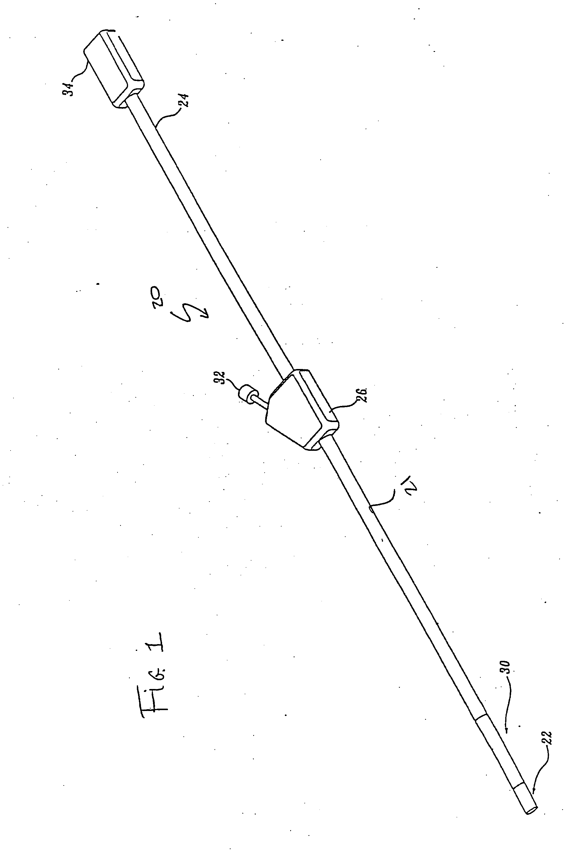
[0024] Generally described, the present invention provides an articulation joint and a method of making an articulation joint for use in a medical device, such as an endoscope. The present invention provides many advantages over articulation joints used in conventional endoscopy systems. For example, the articulation joints of the present invention are easy to assemble and do not require the use of adhesives or brazing, thereby providing an inexpensive and easily mass-produced joint that allows the distal end of a medical device, such as an endoscope, to be bent in any desired direction by one or more control cables.

[0025] The various embodiments of the articulation joint described herein may be used with both conventional reusable endoscopes and low cost, disposable endoscopes, such as those described in U.S. patent application Ser. No. 10 / 811,781, filed Mar. 29, 2004, and in a U.S. Continuation-in-Part patent application Ser. No. 10 / 956,007, filed Sep. 30, 2004, that are assigned...


PUM

PropertyMeasurementUnit
circumferenceaaaaaaaaaa
closing angleaaaaaaaaaa
circumferenceaaaaaaaaaa

Abstract

In one aspect, the present invention is an articulation joint for use in a low cost medical device such as an endoscope intended for a single use. In one embodiment, the articulation joint comprises a plurality of interconnected segments. In another embodiment, the articulation joint comprises an elongated tubular body. In another aspect, the present invention provides methods of manufacturing an articulation joint for use in a medical device.

Description

FIELD OF THE INVENTION [0001] The present invention relates to medical devices in general and to articulation joints for use in medical devices in particular. BACKGROUND OF THE INVENTION [0002] It has become well established that there are major health benefits from regular endoscopic examinations of a patient's internal structures such as the alimentary canals and airways, e.g., the esophagus, stomach, lungs, colon, uterus, urethra, kidney, and other organ systems. Endoscopes are also commonly used to perform surgical, therapeutic, diagnostic, or other medical procedures under direct visualization. A conventional imaging endoscope used for such procedures includes an illuminating mechanism such as a fiber optic light guide connected to a proximal source of light, and an imaging means such as an imaging light guide to carry an image to a remote camera, or eye piece, or a miniature video camera within the endoscope itself. In addition, most endoscopes include one or more working chan...

Claims


Application Information

Patent Timeline
Patent Type & Authority Applications(United States)
IPC IPC(8): A61B1/00
CPCA61B1/00103Y10T29/4998A61B1/0055A61B1/0011A61B1/00071A61B2017/003A61B2017/00314A61M25/0138A61B1/0051A61M25/0133A61B2017/00309A61B2017/00305A61B1/005A61B1/0057A61B1/008
Inventor BOULAIS, DENNIS R.
Owner BOSTON SCI SCIMED INC
Learn More