Vascular opening edge eversion methods and apparatuses

a technology of opening edge and aperture, which is applied in the field of methods and apparatuses for closing punctures and apertures, can solve the problems of large number of steps, high rate of post-puncture hemorrhage, and considerable complications

Inactive Publication Date: 2007-03-01
ABBOTT CARDIOVASCULAR
View PDF112 Cites 185 Cited by
  • Summary
  • Abstract
  • Description
  • Claims
  • Application Information

AI Technical Summary

Benefits of technology

[0012] Devices according to the present invention can utilize a contractible or expandable material, for example with memory characteristics, that allow the members of the device to open and engaed spontaneously on a puncture wound of a blood vessel. Members of such devices can have textured gripping surfaces, tissue hooks, or penetrators, to seize the vessel wall and stabilize the device. Such devices can use the spontaneous opening and closing characteristics to seize the edges of the puncture site, and close them, resulting in a complete vascular closure. Such devices can be kept in a contracted or expanded state (high energy state of a memory material) by a delivery sheath and assume its functional, closing form (low energy state of a memory material) when pushed off a delivery sheath.

Problems solved by technology

With conventional methods, the rate of post-puncture hemorrhage is high, which causes considerable complications.
Difficulties with these methods include the large number of steps necessary to deploy the needles, capture the suture, withdraw the suture, tie the knot, and cut the suture.
In addition, the hole in the blood vessel is often widened by insertion of the instrument, and the suture remains intravascularly on the endothelial surface, and thus can be a nidus for thrombus or intravascular mural hyperplasia with later spontaneous and catastrophic closure of the vessel.
Disadvantages to these methods are related to the high likelihood of thrombosis associated with the intravascular plug or anchor, and the presence of collagen or other bioabsorble materials which cause inflammation, activate the clotting cascade, and increase the likelihood of thrombosis, which, in an arterial system, is catastrophic.
These are generally impractical for catheter-based methods.
This also requires a complicated device, and perforation and thrombosis are very real possibilities.
These clip devices are composed of thick semi-rigid material, and can be placed only with a specialized instruments, and because of the rigidity have great potential to injure or cut the blood vessel.
Disadvantages of these clip devices in general include difficulty in retrieving the device if misplaced, excessive manipulation required, the thickness of the clip material which tends to cut or shear the blood vessel, the large forces that must be used to curve the staples and fix the clips, the increased possibility of tearing the blood vessel, and the general lack of control of the forces being applied to the blood vessel.

Method used


Image

  • Vascular opening edge eversion methods and apparatuses
  • Vascular opening edge eversion methods and apparatuses
  • Vascular opening edge eversion methods and apparatuses

Examples


Embodiment Construction

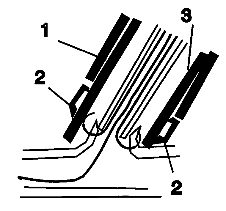
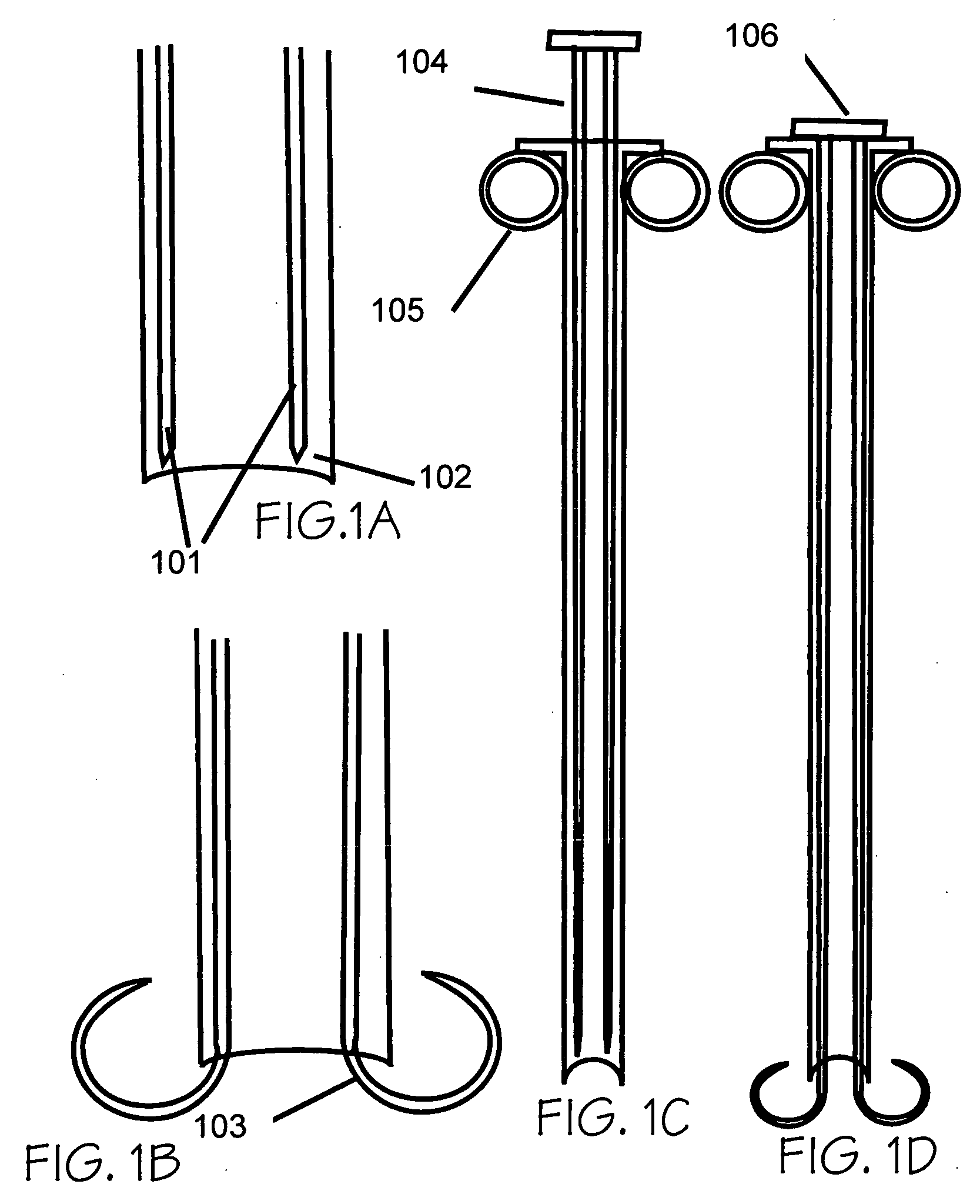
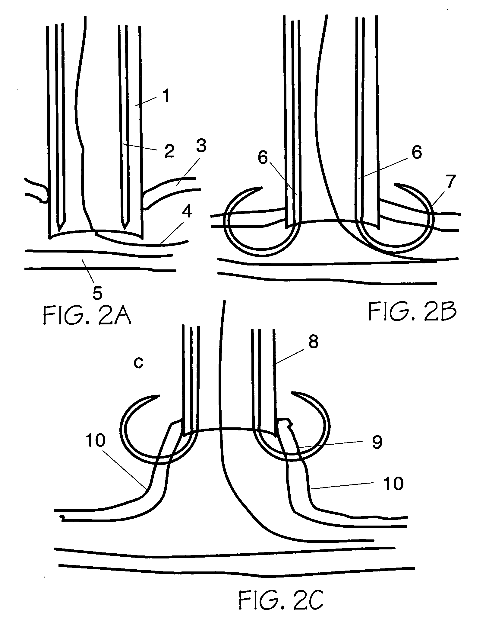
[0023] The present invention provides apparatuses and methods for closing a vascular puncture wound or any tissue aperture, for example those resulting from the insertion of a vascular catheter or surgical instrument, trauma or disease. The present invention embraces both apparatuses and methods for closing tissue openings such as vascular punctures. Devices according to the present invention can be inserted in a vascular sheath, the sheath removed or pulled back, a closure device placed over the everter device, the everted device activated by extending the graspers within the blood vessel, the graspers pulled up against and penetrate the vascular vessel wall, the wound edges everted, apposed, and brought up into the closure device, and finally the everted wound edges closed distal to the graspers by the means of an extravascular clip, extravascular suture, extravascular glue or patch, extravascular heat coagulation, or by staples or sutures that are placed through the lips of the e...


PUM


Abstract

Tissue eversion apparatuses and methods suitable for use, as examples, with vascular closure devices. The tissue eversion apparatuses comprise a tissue engaging element, disposed within a delivery sheath. The delivery sheath protects the tissue engaging element and the surrounding tissue during use. The tissue engaging element can extend from the sheath and engage the tissue near an aperture in a tissue wall. Motion of the tissue engaging element away from the tissue can urge the tissue wall near the aperture to evert from the tissue wall, allowing, as examples, aperture closure apparatuses and methods to be conveniently employed.

Description

CROSS-REFERENCE TO RELATED APPLICATIONS [0001] This application claims priority to U.S. provisional application 60 / 711,279, filed Aug. 24, 2005, and is a continuation-in-part of U.S. utility application 11 / 316,775, filed Dec. 23, 2005, each of which is incorporated herein by reference.FIELD OF THE INVENTION [0002] The present invention relates to methods and apparatuses for closing punctures and apertures in human and animal tissue and to methods and apparatuses for inserting such an apparatus into such tissue to perform such closure functions. BACKGROUND [0003] During angiography and related procedures, catheters are inserted through an incision or puncture in the skin and underlying tissues to access an artery or vein, typically in the groin, neck, or subclavian areas of a patient. The catheter can be inserted through a puncture in the blood vessel and guided to the desired site to perform interventional procedures such as angiography, angioplasty, plaque removal, and infusion of ...

Claims


Application Information

Patent Timeline
Patent Type & Authority Applications(United States)
IPC IPC(8): A61B17/08
CPCA61B2017/00663A61B2017/00004A61B2017/00637A61B17/10A61B2017/00862A61B2017/00672A61B2017/00349A61B17/0057A61B2017/00668
Inventor SIBBITT, WILMER L. JR.SIBBITT, RANDY R.
Owner ABBOTT CARDIOVASCULAR
Why Eureka
Try Eureka