
Sterilization of Biosensors

a biosensor and sterilization technology, applied in the field of sterilization of biosensors, can solve the problems of difficult prediction of the radiation effect on the properties of proteins, and newer, more sophisticated biosensors utilizing pbps or other proteins that require conformational changes for signal transduction, and may be particularly susceptible to denaturation

Inactive Publication Date: 2007-05-17
BECTON DICKINSON & CO
View PDF11 Cites 61 Cited by
  • Summary
  • Abstract
  • Description
  • Claims
  • Application Information

AI Technical Summary

Benefits of technology

The present invention is about methods for making a sterilized biosensor. The methods involve partially assembling the components of the biosensor and separately sterilizing them, followed by aseptically assembling them to produce the sterilized biosensor. The technical effect of the invention is to provide a reliable and efficient way to make a sterilized biosensor.

Problems solved by technology

Radiation effects on the properties of a protein can also be difficult to predict.
Indeed, the newer, more sophisticated biosensors utilizing PBPs or other proteins that require conformational change for signal transduction may be particularly susceptible to denaturation.

Method used


Image

  • Sterilization of Biosensors
  • Sterilization of Biosensors
  • Sterilization of Biosensors

Examples


example 1

Preparation of Alginate Disks and PEG Disks Containing a Binding Protein Entrapped in a Matrix

[0044] A fluorescent-labeled triple mutant of GGBP (“the 3M protein”) was prepared as follows. The 3M protein is a GGBP protein (GenBank Accession No. P02927, without the 23 amino acid leader sequence), and where a cysteine is substituted for an glutamic acid at position 149,an arginine is substituted for an alaninie at position 213 and a serine is substituted for leucine at positiont 238 (E149CA213RL238S). The 3M protein was labeled with IANBD, and the NBD-labeled 3M protein was prepared as described in U.S. application Ser. No. 10 / 040,077, filed Jan. 1 2002, now U.S. Pat. No. 6,855,556 and Ser. No. 11 / 077,028, filed Mar. 1, 2005, and published as U.S. Pre-grant Publication 2005 / 0239155 both of which are incorporated herein by reference.

[0045] Alginate disk were prepared in the following manner. A mix of 2% Alginate in sterile water by weight was prepared. To this solution we added 0.1 M...

example 2

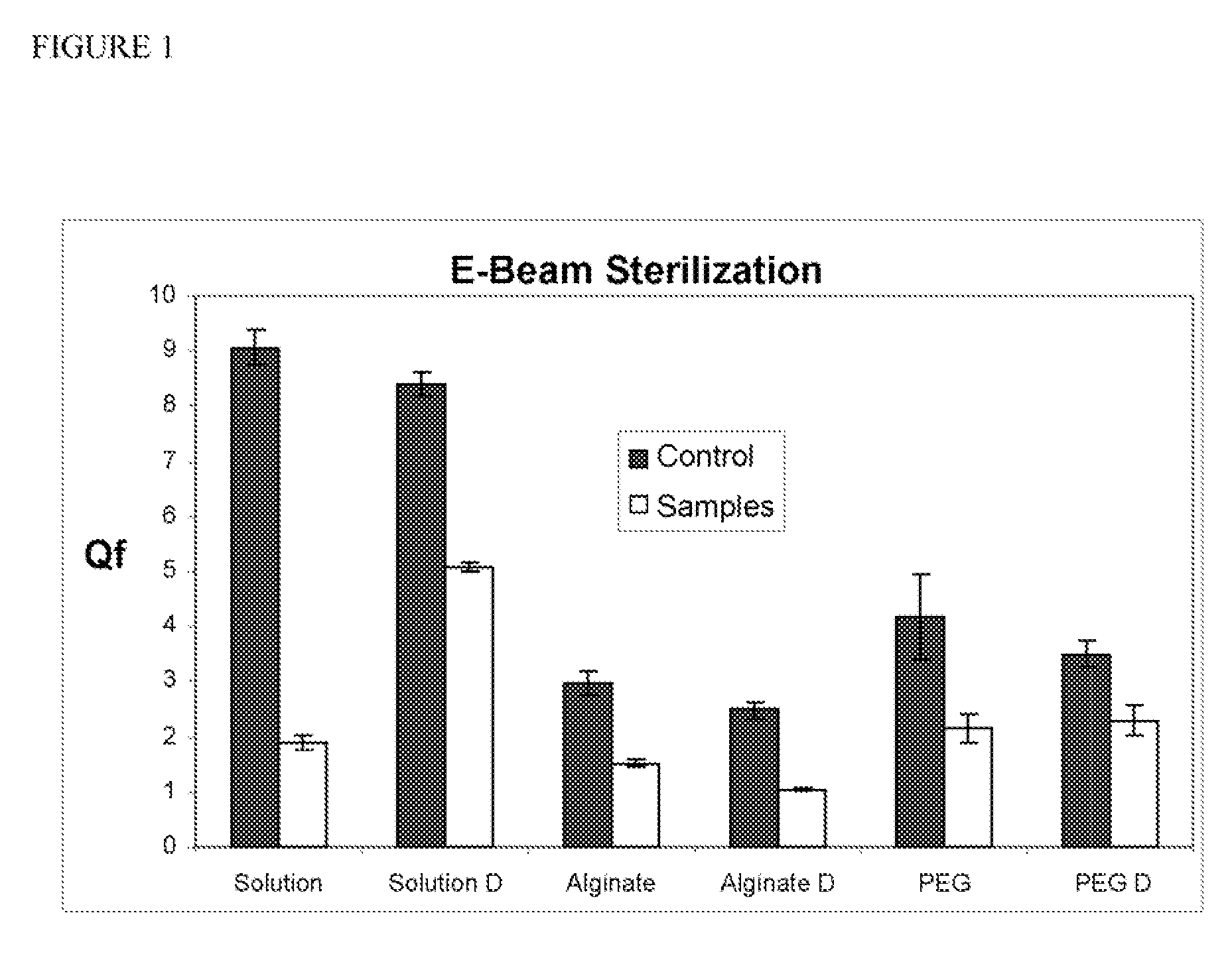
Electron-Beam Sterilization of Non-lyophilized and Lyophilized Disks as Prepared in Example 1

[0049] The wet and dry disks of Example 1 were sterilized using electron-beam radiation. In addition, protein in solution and lyophilized protein were also irradiated using electron-beam radiation. In this experiment, the 20 kiloGrays (2 Mrads) (6.25 kGy / sec) were used, and the dose was confirmed by dosimeter.

example 3

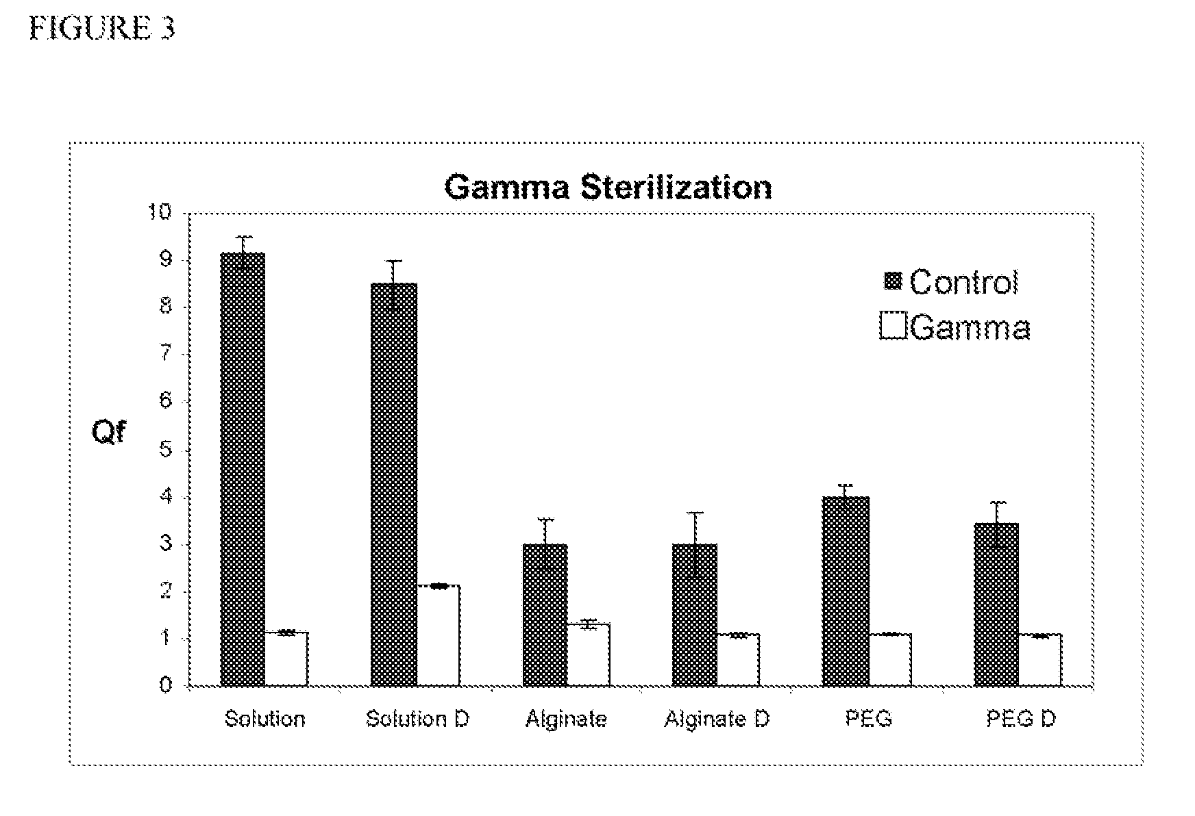
Gamma Radiation Sterilization of Non-Lyophilized and Lyophilized Disks as Prepared in Example 1

[0050] The wet and dry disks of Example 1 were sterilized using gamma radiation. In addition, lyophilized and non-lyophilized protein in solution was also irradiated using gamma radiation. In this experiment, the 20 kiloGrays (2 Mrads) was used. In this experiment, the 20 kiloGrays (2 Mrads) (8.33 kGy / hr) were used, and the dose was confirmed by dosimeter.


PUM

PropertyMeasurementUnit
concentrationsaaaaaaaaaa
pHaaaaaaaaaa
pHaaaaaaaaaa

Abstract

The present invention relates to methods of making a sterilized biosensor, where the biosensor comprises at least one binding reagent, which comprises at least one non-enzyme proteinaceous binding domain. Certain embodiments of the methods described herein comprise partially assembling the components of the biosensor, except for the binding reagent, and separately sterilizing this partial assemblage and the binding reagent. The sterilized binding reagent and the sterilized partial assemblage are then aseptically assembled to produce the sterilized biosensor. Other embodiments of the methods described herein comprise assembling substantially all of the components of the biosensor, including the binding reagent, and sterilizing the assembled biosensor to produce a sterilized biosensor.

Description

CROSS-REFERENCE TO RELATED APPLICATIONS [0001] The application claims priority to U.S. Provisional Application No. 60 / 595,942, filed Aug. 19, 2005, the entire contents of which are incorporated by reference herein.BACKGROUND OF THE INVENTION [0002] 1. Field of the Invention [0003] The present invention relates to methods of making a sterilized biosensor, where the biosensor comprises at least one binding reagent, which comprises at least one non-enzyme proteinaceous binding domain. [0004] 2. Background of the Invention [0005] A variety of implantable electrochemical sensors have been developed for detecting and / or quantifying specific agents or compositions in a patient's blood. For instance, glucose sensors are being developed for use in obtaining an indication of blood glucose levels in a diabetic patient. Such readings are useful in monitoring and / or adjusting a treatment regimen which typically includes the regular administration of insulin to the patient. A rapidly advancing ar...

Claims


Application Information

Patent Timeline
Patent Type & Authority Applications(United States)
IPC IPC(8): C12Q1/00
CPCA61B5/1486A61L2/0011A61L2/0035A61L2/0094A61L2/022A61L2/081A61L2/082A61L2/087A61L2/10A61L2/206A61L2/208
Inventor ALARCON, JAVIERWEIDEMAIER, KRISTINKURISKO, DAVID M.SRIDHARAN, SRINIVASAN
Owner BECTON DICKINSON & CO
Learn More