Method for diagnosis of tuberculosis by smear microscopy, culture and polymerase chain reaction using processed clinical samples and kit thereof

a tuberculosis and smear microscopy technology, applied in the field of multi-purpose diagnostic methodologies for bacterial infections including tuberculosis, can solve the problems of lack of sensitivity, delay of 46 weeks in arriving at a definitive diagnosis, and unfavorable the action of conventional antibiotics and lytic procedures

Inactive Publication Date: 2007-05-17
DEPT OF BIOTECHNOLOGY MINIST OF SCI & TECH GOVERNMENT OF INDIA
View PDF3 Cites 12 Cited by
  • Summary
  • Abstract
  • Description
  • Claims
  • Application Information

AI Technical Summary

Benefits of technology

[0051] The main object of the present invention is to develop an effective and economical method of processing clinical samples useful for simple, rapid, safe, sensitive, and accurate diagnosis of tuberculosis and certain gram positive bacterial infections.

Problems solved by technology

This organism is characterized by its slow growth and the presence of a unique lipid-rich cell wall that makes it unamenable to the action of conventional antibiotics and lytic procedures.
However it suffers from lack of sensitivity since a load of ˜10,000 organisms / ml of specimen is required to give a positive report by Ziehl Neelsen staining from concentrated samples (Kent and Kubica, 1995).
Culture is considered as the gold standard, but on account of the slow growth rate of the pathogen, sole dependence on it would invariably lead to a delay of 46 weeks in arriving at a definitive diagnosis.
Apart from pulmonary tuberculosis, extra pulmonary tuberculosis presents an enormous diagnostic challenge on account of the wide spectrum of organs involved and the paucibacillary load of bacteria in the sample sent to the diagnostic laboratory.
These approaches have suffered in no small measure from the inability to isolate mycobacterial PCR-amplifiable DNA of adequate quality from all types of clinical material.
This presents a very practical and serious impediment to the application of DNA amplification methodologies to clinical material.
NALC and DTT are effective mucolytic agents but are costly.
Zephiran requires neutralization with lecithin and is not compatible with inoculation on egg-based culture media.
For culturing of M. tuberculosis a separate protocol is prescribed wherein three lytic enzymes are used in conjunction with CB-18™ which increases the cost.
It is to be noted that sample processing by this method is not compatible with culturing in liquid media.
But this needs an overnight precipitation and is not suitable for culturing as it kills the mycobacteria.
However the fluorescence microscopy technique would increase the cost.
But with these automated systems it is not possible to study the colony morphology of the cells growing and the absence of contaminants have to be confirmed by performing AFB staining of the positive cultures.
These systems are very costly and out of place in disease-endemic countries like India.
The radiometric detection is a biological hazard too.
But a single method that is (a) compatible with smear microscopy, culture and DNA isolation from clinical samples and (b) suitable for all kinds of pulmonary and extrapulmonary samples, is not available.
The method is cumbersome, involves toxic GITC, tissue processing takes longer time and requires enzymatic digestion, and has been developed only for DNA isolation (Noordhoek et al.
The method though simple, rapid and efficient uses toxic reagent GITC, requires NALC treatment and was not assessed for compatibility with smear microscopy and culture (Chakravorty and Tyagi, 2001).
In summary, these commercial technologies are very sophisticated, costly including high running costs and generally not suitable for developing countries.
This needs proper training of technicians to identify the purulent portion, failure to collect which may result in false-negative smear reporting even from sputum samples containing >10,000 AFB / ml.
Since the purulent portion of the sputum is dealt with, there are chances that the smear may be too thick which is not suitable for smear microscopy.
Even with properly smeared slides, the counterstaining material (due to the mucus, protein and tissue / cell debris present in the sputum) often hinders proper reading of slides).
The process though deals with the entire sputum sample, involves a costly reagent NALC.
Only one or two loopfuls of the concentrated sample is smeared which does not always allow detectable levels of AFB to be present in the smear in case of paucibacillary samples.
Failure to do so renders the sample unsuitable for culturing.
Decontamination using 2% NaOH is not always suitable for tropical countries with a humid climate where use of 3% NaOH is recommended which minimizes the recovery of live mycobacteria due to toxicity effects (Krasnow and Wayne, 1966).
Majority of the methods of DNA isolation, either involve hazardous organic reagents or expensive enzymes.
There is no environment-friendly method that guarantees the isolation of PCR quality inhibitor-free mycobacterial DNA from all types of clinical samples.

Method used


Image

  • Method for diagnosis of tuberculosis by smear microscopy, culture and polymerase chain reaction using processed clinical samples and kit thereof
  • Method for diagnosis of tuberculosis by smear microscopy, culture and polymerase chain reaction using processed clinical samples and kit thereof
  • Method for diagnosis of tuberculosis by smear microscopy, culture and polymerase chain reaction using processed clinical samples and kit thereof

Examples


Embodiment Construction

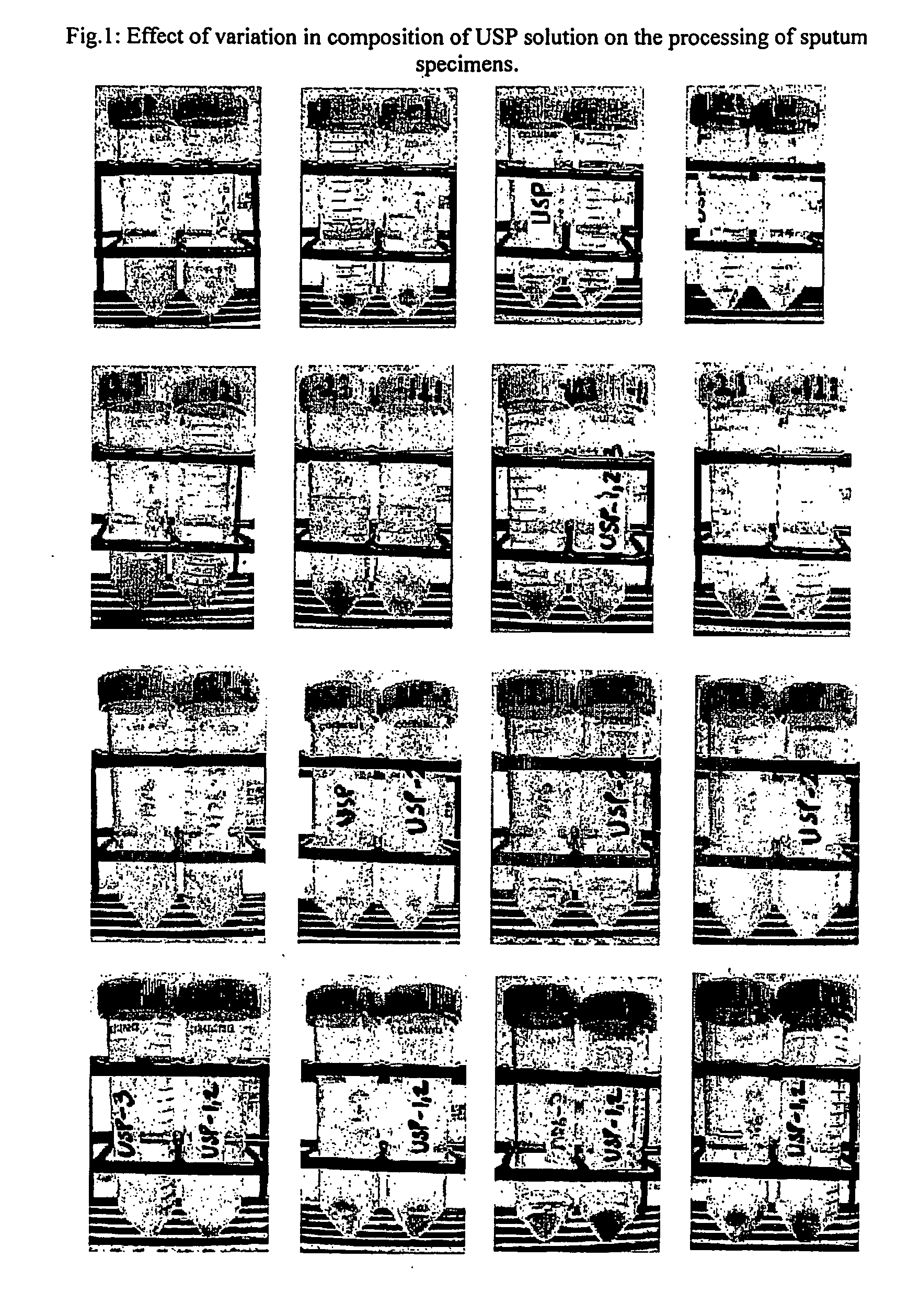
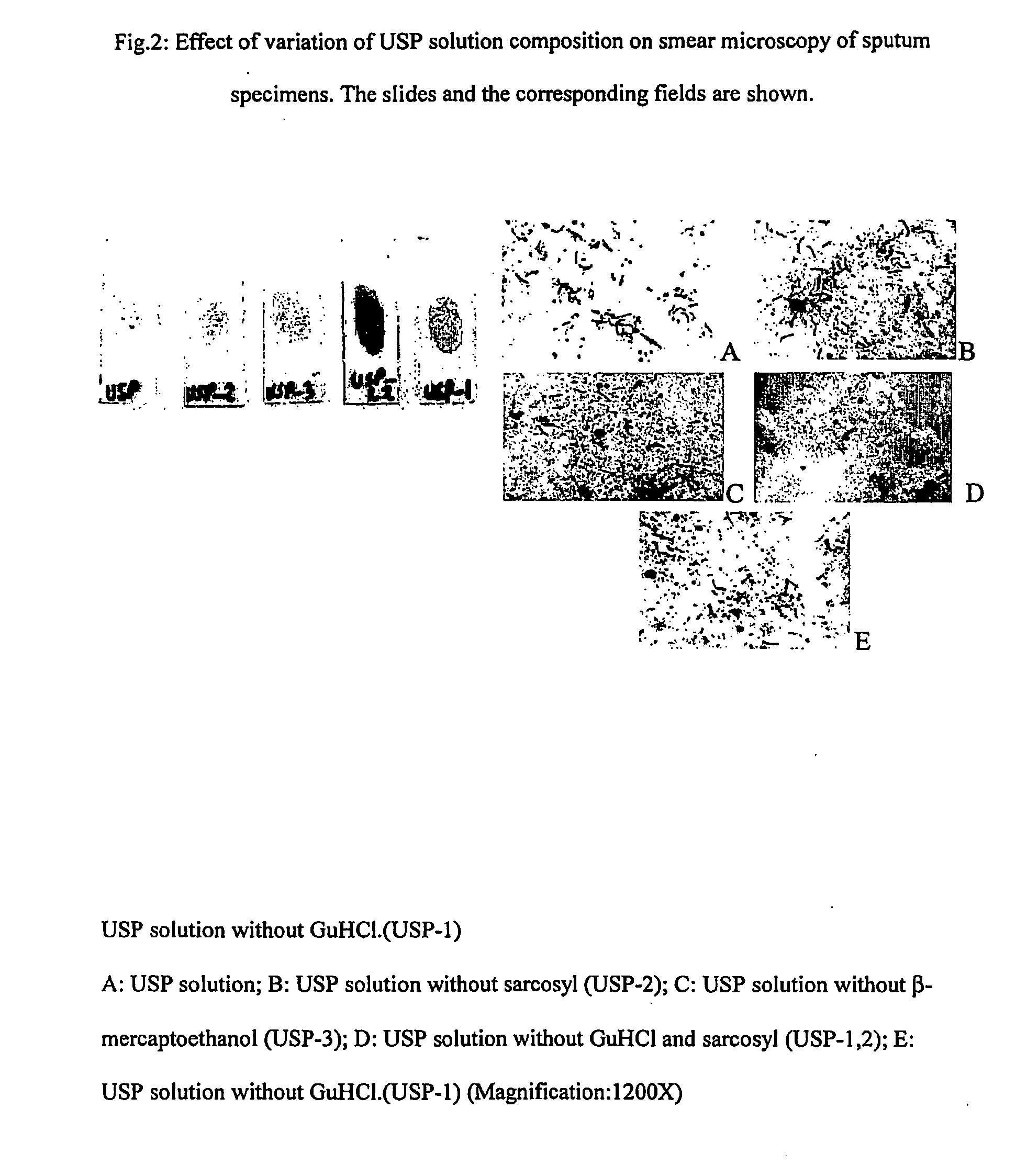
[0070] Accordingly, the present invention relates to an effective and economical method of processing clinical samples useful for simple, rapid, safe, sensitive, and accurate diagnosis of bacterial infections using a composition comprising solution 1 consisting of Universal Sample Processing (USP) solution (composed of Guanidinium Hydrochloride (GuHCl) of concentration ranging between 3-6M, Tris-Cl of concentration ranging between 40-60 mM of pH ranging between 7.3-7.7, EDTA of concentration ranging between 20-30 mM, Sarcosyl of concentration ranging between 0.3 -0.8%, beta-mercaptoethanol of concentration ranging between 0.1-0.3 M), Solution 2 consisting of Sodium phosphate of concentration ranging between 65 to 70 mM of pH ranging between 6.7 to 6.8 (optional can be replaced with sterile water), and Solution 3 consisting of Tween 80 of concentration ranging between 0.03 to 0.08%, and Solution A comprising Chelex 100 suspension of concentration ranging between 8-12%, Solution B con...


PUM

PropertyMeasurementUnit
time durationaaaaaaaaaa
volumesaaaaaaaaaa

Abstract

The present invention relates to a method of processing clinical samples for diagnosing bacterial infections by smear microscopy, culture or PCR as well as a kit for performing the said method. This invention also pertains to two sets of primers specific for the DevR gene of M. tuberculosis as well as to a method of using said primers for screening patients for tuberculosis by PCR.

Description

FIELD OF THE PRESENT INVENTION [0001] A multipurpose diagnostic methodology for bacterial infections including tuberculosis is described that includes processing of clinical specimens and the utilization of the processed material for smear microscopy, culture and polymerase chain reaction. It is applicable for all types of pulmonary and extra pulmonary tuberculosis. BACKGROUND AND PRIOR ART REFERENCES TO THE PRESENT INVENTION [0002] Tuberculosis kills more people in India and South-East Asia than any other infectious disease—more than HIV, STD, malaria and tropical diseases combined. In India more than one thousand people die from TB everyday—more than 450,000 per year, 1 every minute. Rapid diagnosis of tuberculosis is becoming increasingly important to improve cure rates, reduce the risk of spreading the disease through pulmonary route, and to combat the recent emergence of drug resistant strains of M. tuberculosis bacteria and its severe implications in HIV-infected patients. The...

Claims


Application Information

Patent Timeline
Patent Type & Authority Applications(United States)
IPC IPC(8): C12Q1/68C12N1/08
CPCC12Q1/689
Inventor TYAGI, JAYA SIVASWAMICHAKRAVORTY, SOUMITESH
Owner DEPT OF BIOTECHNOLOGY MINIST OF SCI & TECH GOVERNMENT OF INDIA
Why Eureka
Try Eureka