

Method for detection of colorectal cancer in human samples
a colorectal cancer and human sample technology, applied in the field of human sample colorectal cancer diagnosis, can solve the problem of unmet need for a simple diagnostic and/or prognostic tes
- Summary
- Abstract
- Description
- Claims
- Application Information
AI Technical Summary
Benefits of technology
Problems solved by technology
Method used
Image

Examples
example 1
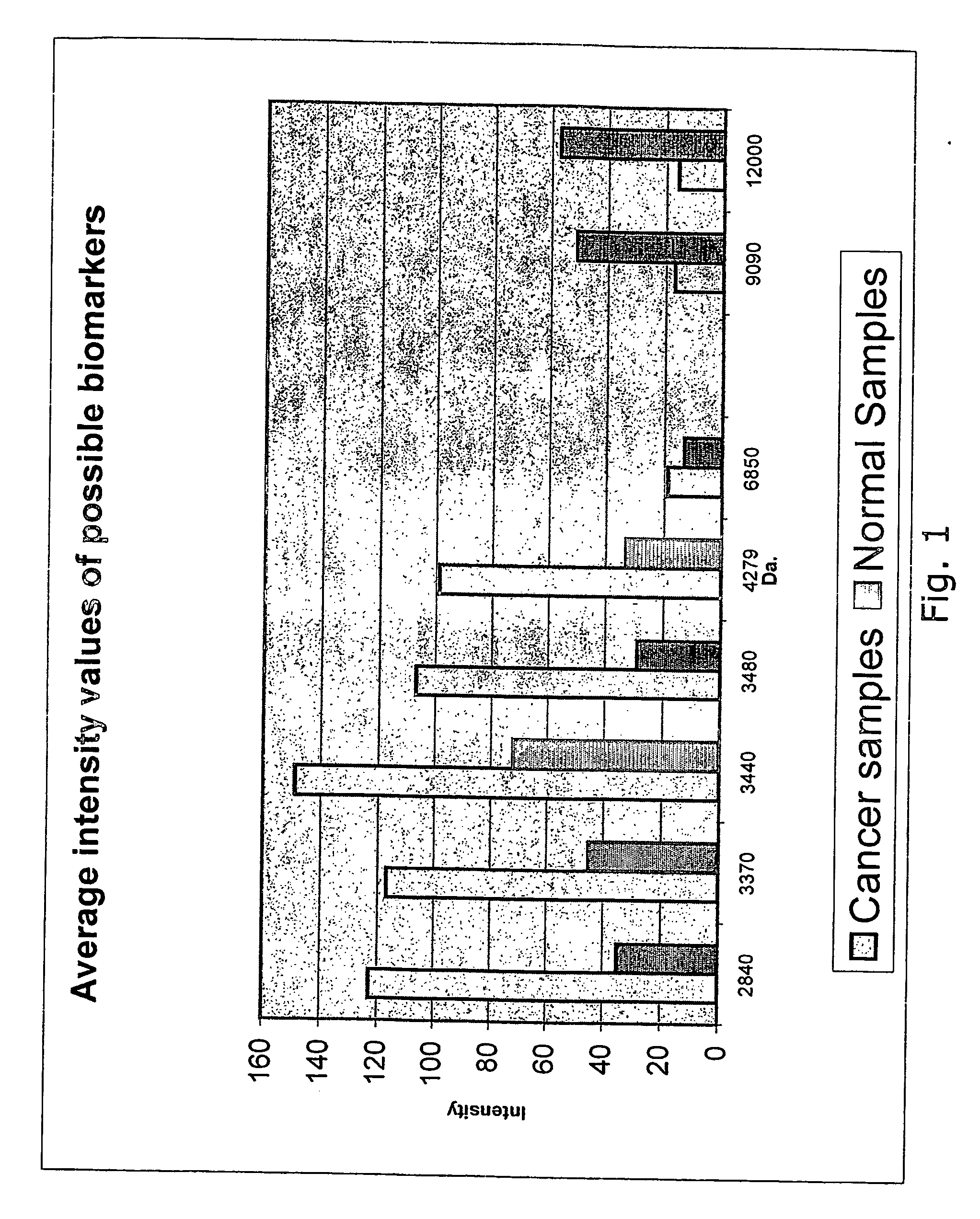
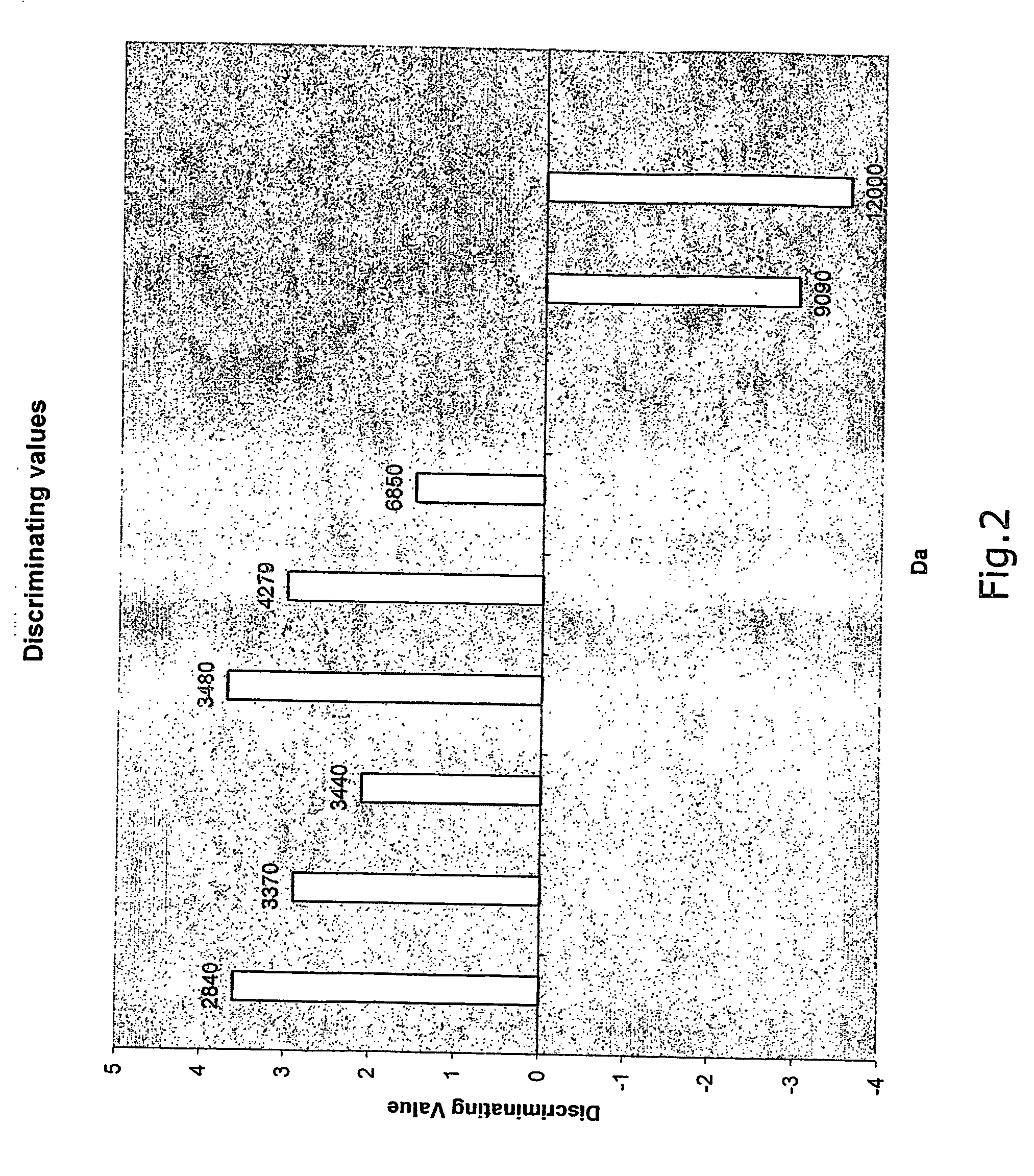
Identification of Biomarkers for Colorectal Cancer by Tissue Investigations
[0186] The aim of the study was to identify protein markers indicative of colorectal cancer by comparison of normal and cancer tissue from colon and rectum.
[0187] Method
[0188] Sample Preparation
[0189] Samples from 12 cancer patients were collected. Normal tissue samples and cancer tissue samples from the same colon were taken and frozen at −80° C. Prior to analysis the samples were taken out of the freezer and placed into homogenisation / Lysis buffer.
[0190] Lysis Buffer: [0191] 100 mM TRIS, pH 8.0 [0192] 9.5 M UREA [0193] 1% CHAPS.
[0194] The samples were homogenised in a Wheaton Overhead Stirrer for 2 minutes at speed step 2.
[0195] Analysis
[0196] Protein extracts were analysed by mass-spectrometry using the SELDI-TOF technique.
[0197] SAX2 chips were pre-treated with 50 μl 100 mM TRIS pH 8.0 buffer.
[0198] 10 μl homogenised sample+60 μl TRIS pH 8.0 buffer were mixed and incubated on SAX2 Chip in a Biop...
example 2
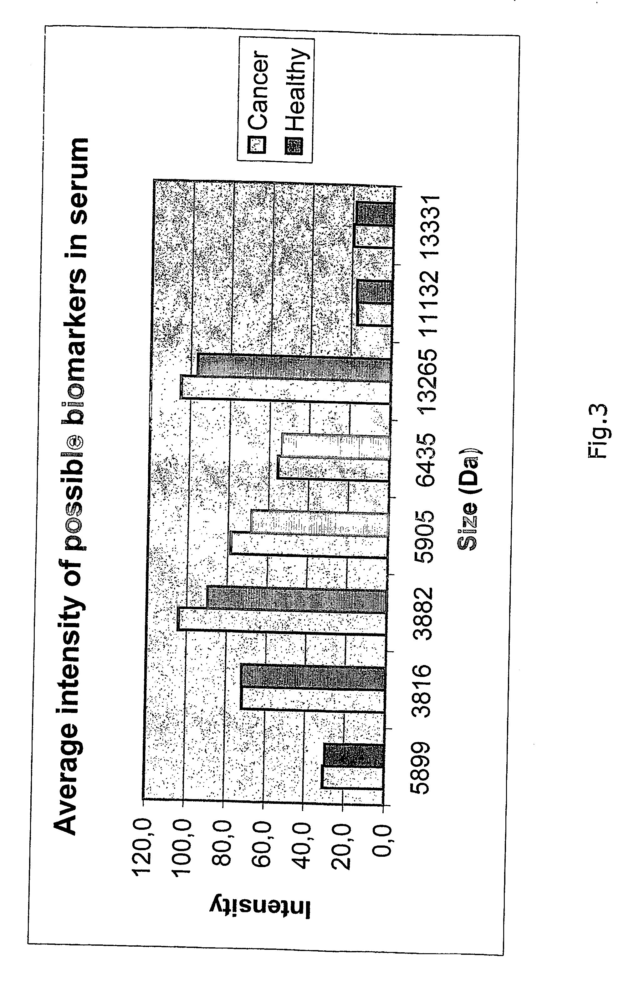
Identification of Biomarkers for Colorectal Cancer in Serum
[0213] The aim of the study was to identify protein markers indicative of colorectal cancer by comparison of serum samples from normal and cancer patients.
[0214] Method
[0215] Sample Preparation
[0216] Serum was isolated from blood of 10 patients diagnosed as having colorectal cancer and 10 healthy individuals.
[0217] Analysis
[0218] An IMAC3 chip was pre-treated with 2 times 5 μl 100 mM NiSO4 followed by wash with 5 μl MQ water and equilibration with 2 times 5 μl binding buffer.
[0219] Binding Buffer: [0220] 100 mM TRIS HCl, pH 7.5 [0221] 500 mM NaCl [0222] 0.1% Triton X-100
[0223] 2 μl of each serum sample was diluted in 48 μl binding buffer of which 4 μl was applied to the protein chip surface. The chip was left on shaker at room temperature for 40 minutes. The sample was removed from the chip surface and each spot was washed with 3 times 5 μl washing buffer (PBS, pH 7.4, 700 mM NaCl). Finally the chip was air-dried and...
example 4
Use of Seldi-TOF / MS or Maldi-TOF / MS for Detection of Biomarkers for Colorectal Cancer.
[0268] The aim of this study was to compare the outcome of markers detected with different expression of proteins in healthy individuals vs. patients diagnosed with colorectal cancer, using either SELDI-TOF / MS or an MALDI-TOF / MS.
[0269] Method
[0270] The PBS II instrument allows variation of three important parameters when analysing protein chips or MALDI-TOF / MS samples.
[0271] Laser intensity, detector sensitivity and optimisation range.
[0272] Laser intensity was permanently set at 220. However, since the laser source is constantly becoming weaker as the instrument is being used, and varies significantly from instrument to instrument, this is not a value that has any general meaning. Most often values from 190 to 230 are chosen.
[0273] Detector sensitivity was set at values of 3, 4, 5, 6, 7, 8 depending on the signal. The intensity (and only the intensity, not the protein profile) of the sample ...
PUM
 Abstract
Description
Claims
Application Information